Preparation and characterization on the SIS/PAA/LAP wound dressing
The LAP nanoparticles owned particle dimension of roughly 10 nm. TEM photos confirmed particular parts equivalent to Mg, Si, Na, and O (Fig. S1). We firstly investigated the gelation properties of the LAP nanoparticles primarily based on electrostatic interactions. As proven in Fig. S2, when the LAP concentrations in water had been low (e.g., 2 wt.% and 4 wt.%), the electrostatic interactions between LAP nanoparticles had been weak to make gel formation troublesome. Because the focus elevated, gel formation turned evident, and the gelation time decreased with greater nanoparticle concentrations. When the LAP focus reached 10 wt.%, the gelation time declined to 86.75 ± 6.70 s. Then, we examined the tissue shear adhesion of PAA hydrogels shaped by cross-linking with totally different concentrations of AA, in keeping with ASTM F2255. The outcomes confirmed that with growing AA focus, the shear adhesion power elevated accordingly. At 40 wt.%, the adhesion reached 23.59 ± 5.59 kPa. Contemplating the upper adhesion power, we selected a 40 wt.% focus of AA for additional experiments.
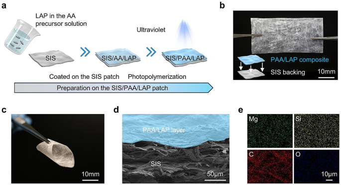
Determine 1a illustrated the schematic strategy of making ready the uneven SIS-based wound dressing, which concerned coating the SIS floor with AA pre-polymerized resolution containing LAP nanoparticles, after which subjected it to UV mild publicity, ensuing within the formation of SIS/PAA/LAP patch. Determine 1b confirmed the bodily look of the SIS/PAA/LAP patch, by estimating its thickness, the PAA/LAP layer owned 4.93 ± 0.67 μm, demonstrating that the PAA adhered to the SIS floor with out affecting the patch’s morphology, and the SIS/PAA/LAP patch exhibited good flexibility (Fig. 1c). SEM examination of the SIS/PAA/LAP revealed that the PAA/LAP coating adhered uniformly to the SIS floor (Fig. S4, Fig. 1d). Moreover, particular aspect mappings of LAP, equivalent to Mg and Si, confirmed the LAP even distribution throughout the SIS/PAA/LAP patch. We additionally performed mechanical assessments on the SIS, SIS/PAA, and SIS/PAA/LAP patches. The outcomes confirmed that the introduction of PAA and PAA/LAP coatings improved the mechanical power. Younger’s modulus and tensile power of SIS patches elevated from 215.15 ± 13.90 MPa and 17.56 ± 3.82 MPa to 360.63 ± 24.37 MPa and 48.90 ± 6.80 MPa, respectively, within the case of SIS/PAA/LAP. This uneven SIS/PAA/LAP construction exhibited tissue adhesion power, as proven in Fig. S6. These outcomes demonstrated that via collection of LAP and AA concentrations, we had been capable of put together asymmetrically adhesive SIS-based wound dressings.
Preparation on the SIS/PAA/LAP wound dressing. a Schematic illustration on fabricating the SIS/PAA/LAP patch, ranging from the AA pre-polymerized resolution containing LAP nanoparticles coated on the SIS patch, to photopolymerization and crosslinking to type PAA/LAP adhesive layer on the SIS substrate. b Gross look on the SIS/PAA/LAP patch. c Flexibility of the SIS/PAA/LAP patch. d Cross-sectioned SEM picture on the SIS/PAA/LAP patch. e Elemental mappings on the SIS/PAA/LAP.
Analysis on the tissue adhesion
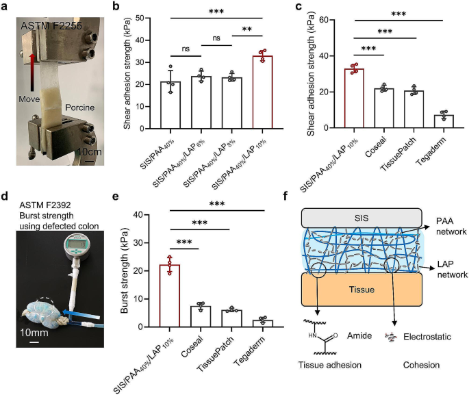
We performed systematic testing to guage the tissue adhesion of the wound dressings. In tissue shear adhesion assessments, when the LAP focus reached 10 wt.%, there was a big improve in shear adhesion power, rising from an preliminary worth of 21.38 ± 4.94 kPa (SIS/PAA40%) to 32.98 ± 2.31 kPa (SIS/PAA40%/LAP10%) (Fig. 2(a, b)), which was attributed to the formation of a second crosslinking community and cohesion beside the PAA community. Moreover, we in contrast the adhesion power of the ready SIS/PAA/LAP with industrial tissue adhesive merchandise, demonstrating that its adhesion power surpassed merchandise equivalent to Coseal (22.05 ± 1.65 kPa), TissuePatch (20.70 ± 2.12 kPa), and Tegaderm (7.25 ± 2.08 kPa). Moreover, we examined the burst power in keeping with the ASTM F2392 customary. The outcomes revealed that the SIS/PAA/LAP had burst power of twenty-two.25 ± 2.50 kPa, considerably greater than Coseal (7.53 ± 1.22 kPa), TissuePatch (6.10 ± 0.62 kPa), and Tegaderm (2.50 ± 0.95 kPa) (Fig. 2(d, e)). We assumed that the mixture of double community buildings together with LAP electrostatic interplay because the second cohesion, and PAA chemical crosslinking hydrogel because the tissue adhesion and first cohesion layers considerably enhanced the fabric’s mechanical power and adhesion power (Fig. 2f) [20, 21]. Consequently, the ready SIS/PAA/LAP wound dressing exhibited glorious tissue adhesion, doubtlessly facilitating wound closure and stopping points such because the detachment of dressings from the wound web site. For ease to explain the totally different teams, within the following assays we remarked SIS/PAA40% and SIS/PAA40%/LAP10% as SIS/PAA and SIS/PAA/LAP to conduct assays.
Tissue adhesion on the SIS/PAA/LAP wound dressing. a Lap shear adhesion assay primarily based on ASTM F2255. b Shear adhesion power with totally different LAP concentrations within the PAA adhesive layers. c Comparisons of adhesion power amongst varied industrial merchandise to the SIS/PAA/LAP. d Burst power assay primarily based on ASTM F2392. e Comparisons of burst power amongst varied industrial merchandise to the SIS/PAA/LAP. f Rationalization on the internal double chemical-physical networks to boost mechanical power. N = 4, **P ≤ 0.01, ***P ≤ 0.001
Mobile response to the SIS/PAA/LAP
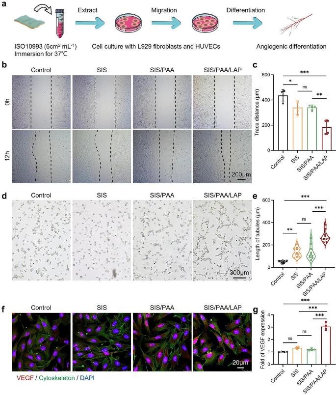
We first performed cytotoxicity analysis following ISO10993 customary by soaking the supplies in cell tradition media for twenty-four hours. The ensuing extraction medium was then used to evaluate the cytotoxicity of the SIS/PAA/LAP on L929 cells and HUVECs (Fig. 3a). As proven in Fig. S7, the outcomes from co-culturing with L929 cells confirmed that the SIS/PAA/LAP teams displayed spindle-shaped cell morphology, indicating that it didn’t induce vital cytotoxicity. Furthermore, with extending tradition interval (as much as 3 days), cell proliferation was noticed, indicating good cell compatibility of the extraction medium. Subsequent, we performed cell scratch, tube formation, and angiogenesis differentiation assays on HUVECs. Determine 3(b, c) confirmed the extracts from the SIS/PAA/LAP considerably promoted HUVEC migration, with a migration price ~ 4.86 occasions than that of the management. Moreover, a modest impact of the SIS in selling cell migration was noticed, presumably as a result of presence of residual bioactive parts equivalent to TGF-β within the materials [14].
Within the tube formation experiment, the SIS/PAA/LAP confirmed denser tube numbers, junction numbers, and longer tube lengths (~ 278 μm), whereas the management owned ~ 50 μm, and the SIS had ~ 133 μm (Fig. 3(d, e)). Within the immunofluorescence staining, the management group confirmed decrease VEGF expressions, whereas within the SIS/PAA/LAP, VEGF expression was ~ 3 occasions than that of the management (Fig. 3(f, g)). These outcomes instructed that the discharge of bioactive ions equivalent to Mg2+ and Si4+ from the SIS/PAA/LAP might intensively improve cell migration and angiogenesis, indicating its potential for efficient tissue restore within the area of sentimental tissue regeneration.
Angiogenesis assays of the SIS/PAA/LAP wound dressing in vitro. a Scheme on making ready the conditioned medium for inducing angiogenic differentiation. b HUVECs scratch assay to estimate cell migration distance. c Semi-quantitative evaluation of hint distance primarily based on panel b. d Tube formation assay of HUVECs after 12-hour incubation. e Semi-quantitative evaluation of tubule size primarily based on panel d. f Immunofluorescent staining of VEGF (purple) /cytoskeleton (inexperienced) / DAPI (blue) after 7-day incubation. g Semi-quantitative evaluation of VEGF fluorescent depth primarily based on panel f. N = 4, *P ≤ 0.05, **P ≤ 0.01, ***P ≤ 0.001
Tissue regeneration within the animal mannequin
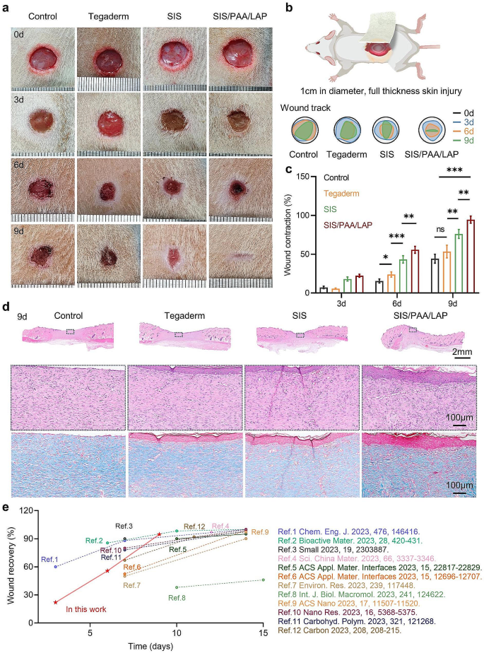
In a full-thickness pores and skin damage mannequin in rats, we initially established round defects with a diameter of 10 mm on the backs of anesthetized rats. Completely different wound dressings (e.g., Tegaderm, SIS, and SIS/PAA/LAP) had been positioned on the wound floor, and wound dressings had been modified each different day following customary care procedures [22]. Upon macroscopic evaluation of the wound therapeutic, the management and Tegaderm teams confirmed poor wound restore, with noticeable wounds after 9 days. The SIS teams had sure wound restore at day-9, whereas the SIS/PAA/LAP teams had largely recovered wounds (Fig. 4a), and an identical tissue restore price was seen within the semi-quantitative wound monitoring evaluation (Fig. 4b). Statistically, postoperative wound contraction at day-6 was 15.45 ± 2.60% for the management teams, 23.88 ± 3.35% for Tegaderm, 43.28 ± 4.79% for SIS, and 55.70 ± 4.42% for SIS/PAA/LAP. At day-9, the wound contraction was 44.20 ± 5.75% for the management group, 53.40 ± 8.17% for Tegaderm, 76.10 ± 5.78% for SIS, and 94.58 ± 4.54% for SIS/PAA/LAP (Fig. 4c).
Combining the previous mobile outcomes (Fig. 3), we supposed that the SIS contained endogenous progress components and different lively substances equivalent to VEGF and TGF-β [14, 23], and the SIS/PAA/LAP enabled firmly adhere to the wound floor. Moreover, the LAP nanoparticles when infiltrated by physique fluids, might launch bioactive ions equivalent to Mg2+ and Si4+, which facilitated tissue therapeutic [24,25,26,27]. Histological staining at 6 and 9 days revealed that the management and Tegaderm teams had apparent wound defects and decrease collagen expression. In distinction, the wound gaps within the SIS, and notably the SIS/PAA/LAP teams turned narrower, with greater collagen mature (Fig. 4d, Fig. S8). This uneven adhesive SIS/PAA/LAP wound dressing with steady ion launch confirmed benefits in wound therapeutic in contrast with different current stories relating to on wound therapeutic supplies (Fig. 4e). Moreover, within the VEGF/DAPI fluorescence staining, the SIS/PAA/LAP teams exhibited greater VEGF fluorescence depth, being 1.84 occasions and a couple of.89 occasions than that of the management group at 6 and 9 days, respectively (Fig. S9). These findings instructed that the ready SIS/PAA/LAP might firmly adhere to wound surfaces and, as LAP launched lively ions, to realize glorious tissue restore outcomes.
In pathological H&E staining of main metabolic organs equivalent to the center, liver, spleen, lung, and kidney at 9 days post-surgery, the tissue morphology and hematological evaluation of the SIS/PAA/LAP teams confirmed no vital distinction in comparison with the management teams, indicating good tissue compatibility (Fig. S10).
Wound regeneration within the rat full-thickness pores and skin damage. a Gross look on the wound restore. b Actual-time wound trajectory on the wound regeneration. c Wound contraction together with time. d H&E and Masson’s trichrome staining on the positioned wound tissues after 9-day. e Our present work in contrast with current wound therapeutic supplies. N = 4, *P ≤ 0.05, **P ≤ 0.01, ***P ≤ 0.001
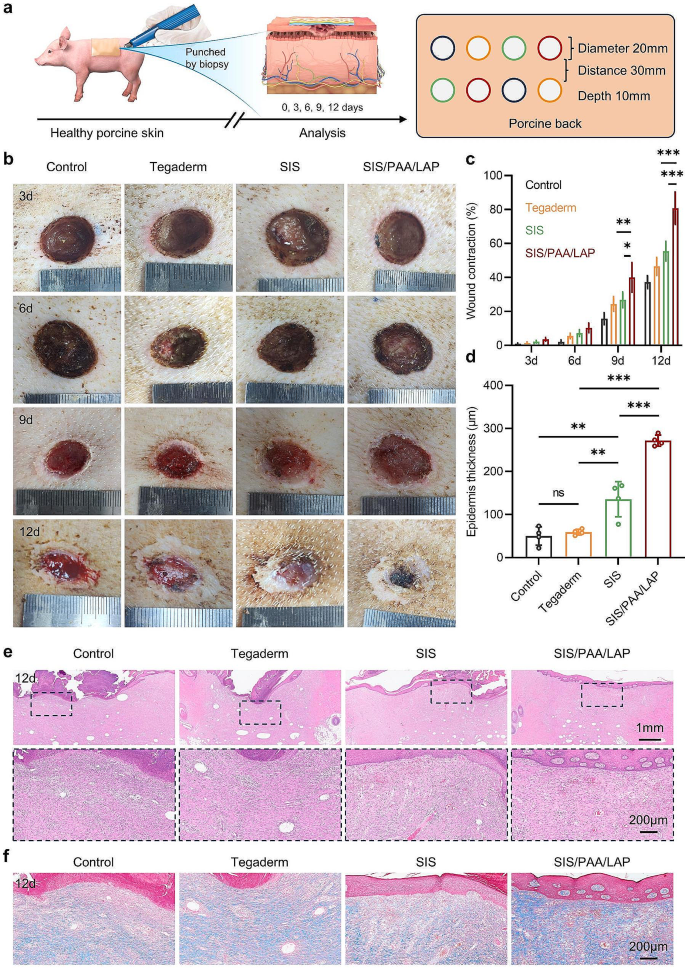
Moreover, in a larger-scale wound defect mannequin on the pores and skin of miniature pig, we evaluated the effectiveness of the wound dressings in selling tissue therapeutic (Fig. 5a). Within the total wound restore final result, all teams exhibited gradual price wound restore within the early stage, nevertheless, after 9 days, the wound restore outcomes turned totally different. At 9 days post-surgery, the SIS/PAA/LAP teams had a wound contraction price of 40.02 ± 8.76%, considerably greater than the opposite teams. At day-12, the wound contraction price of the SIS/PAA/LAP teams reached 80.75 ± 9.53%, whereas the management, Tegaderm, and SIS had charges of solely 37.25 ± 3.77%, 46.50 ± 5.26%, and 55.50 ± 5.80%, respectively (Fig. 5(b, c)). In histological staining, we noticed that the SIS/PAA/LAP wound dressing used on the wound floor didn’t induce antagonistic reactions. Moreover, the usage of SIS and SIS/PAA/LAP led to a big improve in dermis thickness (SIS: 135.63 ± 40.86 μm, SIS/PAA/LAP: 271.75 ± 12.84 μm) in comparison with the management and Tegaderm teams. In Masson’s trichrome staining, the SIS/PAA/LAP teams exhibited greater ranges of collagen expression, with extra orderly collagen association (Fig. 5(d-f)). These outcomes confirmed that the SIS/PAA/LAP had good biocompatibility and was not solely efficient in selling full-thickness pores and skin wound restore in rats but in addition achieved glorious tissue restore ends in a larger-scale miniature pig pores and skin damage mannequin.
Wound regeneration within the Bama miniature porcine pores and skin damage. a Illustration on the experiment routine and pathological evaluation. b Gross look on the wound restore. c Wound contraction together with time. d Dermis thickness progress after 12-day. e H&E staining, and f Masson’s trichrome staining on the positioned wound tissues after 12-day. N = 4, *P ≤ 0.05, **P ≤ 0.01, ***P ≤ 0.001
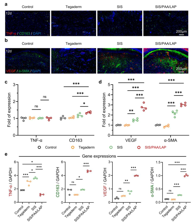
As well as, we performed immunohistochemical staining on the 12-day wound tissue to additional analyze how SIS/PAA/LAP achieved improved tissue restore. We selected TNF-α as a typical pro-inflammatory marker and CD163 as a typical anti-inflammatory marker. Based mostly on the expression of TNF-α/CD163 immunofluorescence, there have been no vital variations in TNF-α expression among the many teams. Nevertheless, the SIS/PAA/LAP teams had 1.31 occasions the depth of CD163 expression in comparison with the management, 1.28 occasions in comparison with Tegaderm, and 1.14 occasions in comparison with SIS (Fig. 6(a, c)). When it comes to VEGF/α-SMA expression, the SIS/PAA/LAP teams exhibited greater expression ranges of VEGF and α-SMA in comparison with the opposite teams. The VEGF was 2.75 occasions than that of the management, 2.79 occasions than that of Tegaderm, whereas α-SMA was 2.99 occasions than that of the management, 3.36 occasions than that of Tegaderm, and 1.41 occasions than that of SIS (Fig. 6(b, d)). And from the RT-PCR evaluation, the precise gene expressions (e.g., TNF-α, CD163, VEGF) had been additionally accorded with the earlier findings (Fig. 6e). These outcomes indicated {that a} vital inflammatory response within the early levels of wound therapeutic [28, 29], when SIS/PAA/LAP was used as a wound dressing, the discharge of bioactive ions equivalent to Mg2+ and Si4+ enabled to extend the expression ranges of intercellular anti-inflammatory components (e.g., CD163), to create a microenvironment conducive to cell migration and proliferation [30, 31]. Moreover, Mg2+ and Si4+ might considerably promote angiogenesis in tissues [27, 32, 33]. Due to this fact, SIS/PAA/LAP was able to attaining glorious tissue wound restore outcomes.
Histological evaluation on the wound regeneration in pig. a Immunofluorescent staining of TNF-α (purple) / CD163 (inexperienced) / DAPI (blue), and b VEGF (purple) / α-SMA (inexperienced) / DAPI (blue) on the positioned wound tissue after 12-day. c Semi-quantitative evaluation of TNF-α / CD163, and d VEGF / α-SMA fluorescent depth expression primarily based on panel a and b, N = 4. e RT-PCR evaluation of the retrieved granulation tissues for particular gene expressions. N = 3, *P ≤ 0.05, **P ≤ 0.01, ***P ≤ 0.001


