Nanostructure of various nanofiber scaffolds
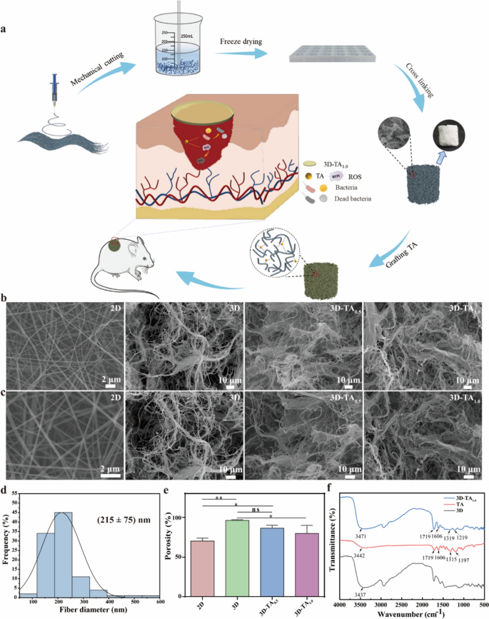
The preparation of 3D-TA is proven in Fig. 1a. Briefly, 2D nanofiber membranes have been fabricated by electrospinning firstly. Then 2D nanofiber membranes have been scissored into items and homogenized by a homogenizer. Secondly, the homogenized nanofibers have been poured into particular molds and freeze-dried for 12 h to arrange 3D nanofiber sponges (3D). The functionalization of 3D nanofiber sponges (3D-TA) was fulfilled by immersing ready 3D nanofiber sponges into TA resolution. The wound therapeutic capability was verified by observing the wound therapeutic means of round full-thickness wounds of mice by protecting with 3D-TA nanofiber sponges.
The macrostructure of bulk 3D nanofiber sponges is proven in Extra file 1: Fig. S1, the place the macrostructure of 3D nanofiber sponges various together with the fabrication mildew and nanofiber sponges with totally different shapes and heights have been ready. The nanostructure of 2D/3D nanofiber scaffolds is proven in Fig. 1b, c. It’s obvious that the 2D nanofiber membrane reveals porous construction with easy nanofiber floor and uniform nanofiber diameter with the distribution of 215 ± 75 nm (Fig. 1d). The morphology of 3D nanofiber scaffolds reveals a definite hierarchical spongy construction with giant dimension pores starting from 32 to 109 μm and quite a lot of small dimension pores starting from 7 to twenty μm. On this hierarchical porous construction, the big pores are fashioned by sublimation of huge ice crystals throughout freeze-drying of the scaffolds that are thought to facilitate cell ingrowth and cell migration throughout the scaffold; Whereas the small pores are self-assembled from tangled quick fibers which facilitate cell adhesion and proliferation [26]. In the meantime, the porous construction can also be conducive to the entry of vitamins and the elimination of metabolic waste. Determine 1e reveals the porosity of various nanofiber scaffolds. The outcomes present the porosity of 2D nanofiber membrane is 70% whereas that of 3D nanofiber sponge is 96%. This greater porosity makes the 3D nanofiber sponge possess greater particular floor space, enhancing the absorption of wound exudates and the trade of gasoline and vitamins successfully. The grafting of various concentrations of TA (3D-TA0.5 and 3D-TA1.0) didn’t have an effect on the hierarchical construction of 3D nanofiber scaffold however barely decreased the porosity and particular floor space. That could be resulting from the truth that with the rise of TA content material on the floor and inside pores of nanofiber sponge, extra hydrogen bonds are fashioned between fibers and TA, leading to adhesion between fibers, thus lowering the porosity and particular floor space of the sponge [38]. Nonetheless, the porosity of 3D-TA0.5 and 3D-TA1.0 nanofiber sponge remains to be greater than that of 2D nanofiber mat with the porosity of 86% and 80%, respectively.
Whether or not the TA was efficiently grafted onto the 3D nanofiber sponges was verified by FTIR (Fig. 1f). The outcomes present that the 3D-TA1.0 nanofiber sponge possesses the attribute peak of carboxylic acid (C = O) vibration at 1719 cm− 1, and the benzene ring C = C vibration absorption peak at 1606 cm− 1 and benzene ring vibration substitution attribute peaks at 1219 cm− 1. These attribute peaks have been in keeping with that of TA indicating the profitable grafting of TA onto the 3D nanofiber sponge. In contrast with the hydroxyl attribute absorption peak of 3D (3437 cm− 1), the hydroxyl attribute peak of 3D-TA1.0 shifted to the left (3471 cm− 1), which attributed to the intermolecular interplay between PVA and TA ensuing within the hydrogen bonding [39]. FTIR spectra confirmed that TA was efficiently grafted onto the 3D scaffold by hydrogen bonds.
Absorption and water retention capability
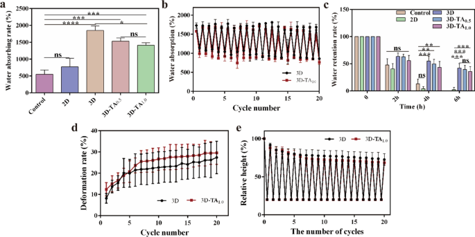
Wound dressings ought to possess good absorption capability of wound exudates to stop an infection [38]. The water absorption capacities of Management, 2D, 3D, 3D-TA0.5 and 3D-TA1.0 are proven in Fig. 2a with ARwater values of 551.4% ± 105.7%, 774.2% ± 209.5%, 1848.4% ± 111.6%, 1533.6% ± 77.0% and 1410.0% ± 64.2%, respectively. It’s obvious that the absorption capability of 3D sponge is perfect amongst all of the samples (1848.4% ± 111.6%), a lot greater than that of Management (551.4% ± 105.7%) and 2D nanofiber membrane (774.2% ± 209.5%). That’s as a result of the microscale layered porous construction of 3D sponge enhances the water entry and penetration capability. Furthermore, the hierarchical community construction and excessive porosity of 3D sponge (96%) present nice water storage capability, making it a superb water absorbent. The water absorption of 3D-TA0.5 and 3D-TA1.0 sponges decreased to 1533.6% ± 77.0% and 1410.0% ± 64.2%, as a result of the grafting of TA decreased the porosity of the 3D sponge (86% and 80%), thus the water absorption of 3D-TA0.5 and 3D-TA1.0 sponges decreased. Nonetheless, the hierarchical community construction was not affected after the grafting of TA, subsequently the water absorption of 3D-TA0.5 and 3D-TA1.0 scaffolds was nonetheless a lot greater than that of Management and 2D nanofiber membranes.
The cyclic water absorption capacities of 3D and 3D-TA1.0 have been verified by a cyclic experiment (Fig. 2b). The outcomes present the 3D and 3D-TA1.0 may retain their exceptional water adsorption capability after repeating twenty adsorption/desorption cycles of water as a result of speedy diffusion and infiltration of water into the scaffolds. When the deformed 3D scaffolds are re-immersed in water, the dehydrated samples will rapidly and spontaneously take up water, stimulating the unfolding of the 3D nanofiber sponges attributable to compression, ensuing within the reversible hygroscopicity of the 3D nanofiber sponges [40].
Wound dressings shouldn’t solely have good efficiency of absorbing exudates, but in addition have the flexibility to keep up a correct moist microenvironment to advertise the regeneration of pores and skin epithelization. The water retention skills of Management, 2D, 3D, 3D-TA0.5 and 3D-TA1.0 inside 6 h is proven in Fig. 2c. The outcomes present the RRwater of Management and 2D teams decreased to 0 inside 6 h whereas that of 3D, 3D-TA0.5 and 3D-TA1.0 nonetheless maintained 42.0% ± 7.0%, 39.4% ± 6.2%, 35.8519% ± 7.4%, respectively. Once more, the water retention capability of 3D is the optimum one. This means the 3D, 3D-TA0.5 and 3D-TA1.0 sponges have good water retention capability due to the wonderful hierarchical porous construction.
Compression/resilience property
The excessive elasticity of wound dressing may keep away from the wound from being squeezed and deformed by the stress of surrounding tissues or washed away by blood stream when dealing with a wound with a sure depth [41, 42]. The restoration capability of 3D nanofiber sponges after compression is proven in Extra file 1: Fig. S2, which signifies the rebound peak of 3D sponge is decrease than the unique peak. The quantitative evaluation of the DRs of 3D and 3D-TA1.0 after twenty repeated compression/resilience experiments is proven in Fig. 2d. It’s discovered that the DRs of each samples enhance constantly to ~ 30% in the course of the preliminary 5 cycles. That’s as a result of the porosity of the 3D samples is giant, and the pattern density is small. When the compression was utilized, the deformation charges of each samples would enhance constantly. After 5 cycles of compression/resilience, each samples have been compressed barely, thus the porosity was decreased and the density of the pattern would enhance. Due to this fact, samples wouldn’t rebound to the unique peak (Fig. 2e). As well as, the DRs are steady after 5 cycles of compression/resilience, indicating that the pattern peak stays unchanged. Evaluating the rebound price of 3D and 3D-TA1.0 samples after twenty cycles of compression/resilience, it may be seen that there’s little distinction between 3D and 3D-TA1.0. Each samples can rebound to 75% of its personal peak after twenty cycles of compression/resilience, which proves that 3D nanofiber sponges possess steady mobile community construction exhibiting good compression/resilience efficiency.
Antioxidant exercise
The antioxidant exercise of wound dressings has been proved to have a constructive impact on the wound therapeutic course of because it regulates the extreme manufacturing of ROS [43]. The antioxidant actions of nanofiber sponges have been evaluated by figuring out their SADPPH. The absorption plots of DPPH free radical options uncovered to totally different samples are proven in Fig. 3a. The absorption curves of 3D and Management group utterly coincide indicating the an identical intensities. Nonetheless, the absorption intensities of 3D-TA0.5 and 3D-TA1.0 are considerably decreased as a result of presence of wierd variety of electrons in DPPH radicals at λ517 nm. When electrons are paired with hydrogen atoms in antioxidants, the absorption depth will scale back, which is able to end result within the colour change of DPPH from purple to yellow (Fig. 3b). The diploma to which the colour turns into lighter is proportional to the variety of electrons eliminated. Determine 3c reveals the relative quantified antioxidant properties of three sorts of sponge samples by analyzing the absorbance change at λ517 nm with ultraviolet spectrophotometer. It’s proven that the SADPPH of 3D, 3D-TA0.5 and 3D-TA1.0 have been 0.0%, 66.2 ± 6.4% and 95.5 ± 0.5%, respectively, which signifies that the grafting of TA makes the 3D nanofibers sponge have apparent antioxidant exercise and that the upper the TA focus, the higher the antioxidant exercise. That’s as a result of TA has phenolic hydroxyl, a powerful free radical terminator, which allows the discount the DPPH free radical to the yellow diphenyl picric acid hydrazine, thus attaining the impact of anti-oxidation [39].
In vitro coagulation efficiency
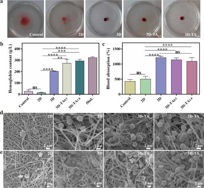
The hemostasis capability is dependent upon how successfully the blood clots on the pattern. The clotting of Management, 2D, 3D, 3D-TA0.5 and 3D-TA1.0 samples are proven in Fig. 4a. It’s noticed that the water within the petri dish turned crimson for the Management and 2D nanofiber membrane, which signifies that the blood is troublesome to clot on medical gauze and 2D fibrous membranes. For 3D, 3D-TA0.5 and 3D-TA1.0 samples, the blood coagulated nicely on the samples, displaying clear water within the petri dish. This means that the 3D nanofiber sponges may speed up blood coagulation when wound is bleeding, and the grafting of TA had no impact on the coagulation impact of 3D nanofiber sponges.
To quantify the hemostatic capability of varied samples, the HCs of Management, 2D, 3D, 3D-TA0.5 and 3D-TA1.0 have been measured (Fig. 4b). The HC of blood (50 mL) was thought of as Management. The upper HC of the examined resolution signifies its higher coagulation impact. The outcomes present the HCs of Management, 2D, 3D, 3D-TA0.5 and 3D-TA1.0 samples have been 26.2 ± 10.3 g/L, 14.4 ± 3.4 g/L, 203.2 ± 2.0 g/L, 273. 0 ± 29.1 g/L, 294.4 ± 11.2 g/L, respectively. It’s obvious that the HCs of 3D, 3D-TA0.5 and 3D-TA1.0 have been a lot greater than that of Management and 2D samples. As a result of the considerable porous construction of 3D, 3D-TA0.5 and 3D-TA1.0 may quickly absorb blood and inhibit the lack of crimson blood cells and platelets from the wound, thus considerably enhancing their hemostatic capability. Moreover, the grafting of TA (3D-TA0.5 and 3D-TA1.0) additional improved the clotting capability of 3D fiber sponges. That’s as a result of polyphenols can kind precipitable complexes with proteins within the blood in a non-specific method to advertise blood clotting [44].
As well as, the ACblood was additionally evaluated as proven in Fig. 4c. The Management and 2D nanofiber membrane teams had poor blood absorption capability with the ACblood worth of 424.8% ± 53.9% and 502.6% ± 74.7%, whereas the 3D, 3D-TA0.5 and 3D-TA1.0 had glorious blood absorption capability with the ACblood worth of 1211.5% ± 38.9%, 1140.6% ± 52.7% and 1089.6% ± 102.9%, respectively. Once more, the extremely porous construction of the 3D nanofiber sponges accelerated the blood absorption. The addition of TA may have an effect on the porosity of the 3D nanofiber scaffolds, thus would additional have an effect on the blood-sucking capability of the 3D nanofiber sponges. Nonetheless, the distinction just isn’t vital.
The interplay between totally different samples (2D, 3D, 3D-TA0.5 and 3D-TA1.0) and blood cells was characterised by SEM (Fig. 4d-e). It demonstrates that only a few blood cells adhere to the 2D nanofiber membrane, whereas numerous blood cells adhere to the 3D, 3D-TA0.5 and 3D-TA1.0 sponges and most of them immerse into the pattern pores, which signifies the hierarchical porous construction of 3D sponges promote the blood coagulation.
Antibacterial properties
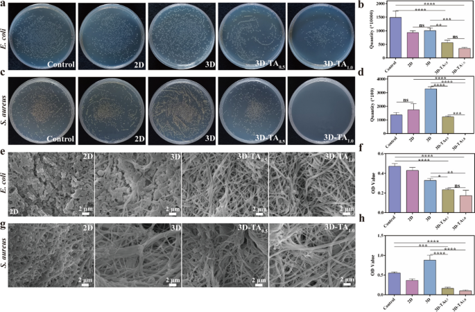
Antibacterial capability is a vital issue for the medical software of wound dressing. The antibacterial actions of Management, 2D, 3D, 3D-TA0.5 and 3D-TA1.0 have been decided by plate counting technique, SEM statement and CCK-8 technique. Determine 5a, c reveals the pictures of the colony-forming of E. coli (Fig. 5a) and S. aureus (Fig. 5c) (106 CFU/mL) on agar plate after being handled with totally different samples. The corresponding colony depend result’s proven in Fig. 5b, d. It demonstrates that the scaffolds grafted with TA (3D-TA0.5 and 3D-TA1.0) have glorious antibacterial efficiency in contrast with the Management, 2D and 3D samples. Furthermore, the bacteriostatic impact enhances with the rise of TA focus as a result of TA can inhibit the expression of toxin genes in micro organism inducing cell membrane injury, thus resulting in the dying of bacterial[45]. As well as, the antibacterial impact of TA on S. aureus is greater than that of E. coli. As a result of E. coli has a further lipopolysaccharide cell wall, which is an efficient barrier in opposition to antibacterial brokers and is extra proof against plant extracts [46]. The antibacterial skills of various samples in opposition to E. coli (Fig. 5e) and S. aureus (Fig. 5g) have been additional verified by SEM. The outcomes are in keeping with that of plate counting technique, indicating higher antibacterial exercise after the grafting of TA onto the 3D nanofiber sponges. Determine 5f, h present the antibacterial capability of various samples in opposition to E. coli (Fig. 5f) and S. aureus (Fig. 5h) through CCK-8 detection technique. The outcomes present the OD worth of 3D-TA0.5 and 3D-TA1.0 have been considerably decrease than these of different teams, which once more demonstrates the wonderful antibacterial capability of 3D nanofiber sponge functionalized with TA.
Antibacterial properties. Images of colony-forming models of a E. coli and c S. aureus on plate after treating with totally different samples and the corresponding colony depend for b E. coli and d S. aureus. SEM photos of e E. coli and g S. aureus on 2D, 3D, 3D-TA0.5 and 3D-TA1.0 surfaces and the antibacterial capability of various samples in opposition to f E. coli and h S. aureus through CCK-8 technique
Cell viability
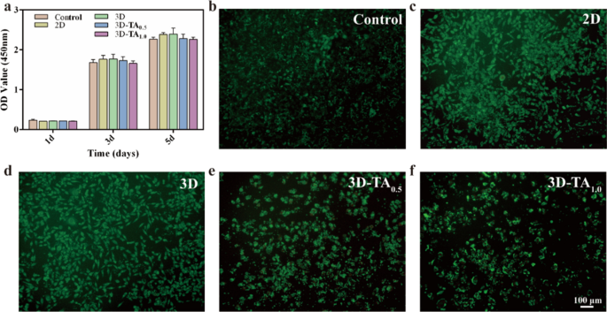
Wound dressings must be extremely biocompatible and promote wound therapeutic via cell proliferation [47]. The cytocompatibility of various dressings was decided by CCK-8 and stay/useless staining experiments. Determine 6a reveals the CCK-8 outcomes of co-culture cells with extracts of various samples for 1, 3, and 5 days. The Management group was pure medium. It may be seen that the OD values of various samples co-cultured cells with totally different extracts for 1 day have been mainly the identical as these of the Management with no vital distinction, which signifies all samples had good biocompatibility. With the rise of co-culture time, the OD values of all options have been elevated, which proved that the 2D, 3D, 3D-TA0.5 and 3D-TA1.0 samples had the flexibility of selling cell proliferation. Though the OD values of 3D-TA0.5 and 3D-TA1.0 have been barely decrease than these of different teams after co-culturing for 3 and 5 days, there was no vital distinction, indicating that the grafting of TA had little impact on cell biocompatibility.
As well as, the live-dead cell experiment was additionally carried out to confirm the great biocompatibility of various samples. Determine 6b–f and Extra file 1: Fig. S3 present the fluorescence photos of L929 cells co-cultured with totally different extracts for 3 days. Right here, numerous residing cells (inexperienced) and only a few useless cells (crimson) are noticed for each the Management and experimental teams (2D, 3D, 3D-TA0.5,3D-TA1.0) after co-culturing for 72 h. The fluorescence photos have been in keeping with these of CCK-8 outcomes, which demonstrated the great biocompatibility of 3D-TA1.0.
In vivo wound closure capability
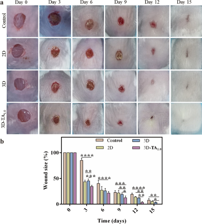
To confirm the mobile ends in vitro, the in vivo wound closure effectivity was evaluated by treating full-thickness wound on the dorsal area of mice with totally different dressings. The medical gauze was used as Management. Determine 7a reveals the optical pictures of wound look after treating with totally different samples (Management, 2D, 3D and 3D-TA1.0) at numerous day intervals (0, 3, 6, 9, 12, 15 days). It’s demonstrated that the wound dimension of all teams decreased with time after remedy. Particularly within the 3D-TA1.0 group, the discount of the wound space was the quickest with an nearly utterly therapeutic on the fifteenth day whereas the opposite three teams coated by Management, 2D and 3D have been a lot bigger than that handled with 3D-TA1.0. The quantitative evaluation of the share of wound dimension over the identical time interval from the pictures (Fig. 7a) is proven in Fig. 7b. The wound dimension of 3D-TA1.0 group was considerably smaller than that of different teams in any respect research time factors. Within the preliminary stage (day 3), the wound space coated by 3D-TA1.0 was decreased to 34.55% ± 1.88% whereas that coated by Management, 2D and 3D was 85.09% ± 3.48%, 43.59% ± 1.06% and 45.33% ± 2.91%, respectively. On the ninth day, the wound space of Management, 2D, 3D and 3D-TA1.0 teams have been 23.06% ± 2.79%, 21.95% ± 3.86%, 19.05% ± 2.18% and 12.64% ± 2.74%, respectively. Additionally it is indicated that the wound closure capability of 3D group was higher than that of Management and 2D group. At 15 days, the wound in 3D group was mainly healed and the wound in 3D-TA1.0 group was utterly healed with out intensive scar and epidermal tissue defect, whereas the apparent wounds have been nonetheless existed within the different experimental teams. The quantitative outcomes have been in keeping with the macroscopic statement outcomes, indicating the efficient enchancment of pores and skin tissue regeneration and promoted cell proliferation capacities of 3D-TA1.0.
Hematoxylin and eosin and Masson’s trichrome
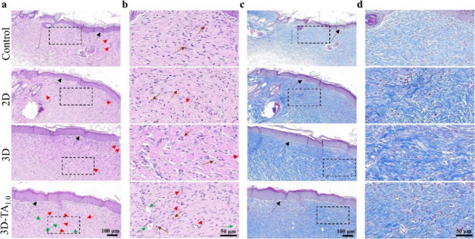
To substantiate the therapeutic impact of varied dressings on wounds, the wound tissues dissected after 15 days of remedy have been subjected to hematoxylin and eosin (H&E) and Masson’s trichrome staining (Fig. 8). The H&E staining (Fig. 8a, b) reveals that the wound after remedy with 3D-TA1.0 underwent extra epidermal tissue regeneration (indicated by black arrows) and the presence of fewer irritation cells (indicated by brown arrows) than that handled with Management, 2D and 3D samples. Furthermore, the 3D-TA1.0 handled wound exhibited extra angiogenesis (indicated by crimson arrows) and hair follicle cells (indicated by inexperienced arrows). The angiogenesis may promote the migration of fibroblasts to the injured website and preserve low ranges of irritation, which ultimately results in a regenerated higher cortex on the wound [48], selling the wound therapeutic[49]. The Masson trichrome staining (Fig. 8c, d) additional verified that extra collagen (blue fibrous construction) was deposited after being handled with 3D-TA1.0 and the association of collagen fibers was extra densely and orderly, much like regular pores and skin [50].
General, the 3D-TA1.0 exhibit glorious wound therapeutic capability, which is attributed to the distinctive hierarchical porous construction of 3D nanofibers sponge facilitating nutrient transport, growing the air permeability, selling fibroblast proliferation throughout wound therapeutic. Furthermore, the TA-functionalized 3D sponge endows the 3D-TA sponge with glorious antioxidant and anti inflammatory properties. The lower of ROS can scale back inflammatory response, scale back the secretion of pro-inflammatory cytokines, enhance the variety of macrophages, and set off the technology of collagen across the wound [51, 52]. As well as, 3D-TA nanofiber sponges additional promote angiogenesis and epidermal tissue regeneration, thus speed up wound therapeutic.


