
(Nanowerk Information) A microscopic shock wave has been photographed passing by way of a single organic cell, due to a brand new pictures method. Nanosecond pictures makes use of ultrafast digital cameras to take pictures on the pace of a billionth of a second. Nonetheless, picture high quality and publicity time are usually restricted. Now, a workforce led by researchers on the College of Tokyo has achieved superfine pictures taken over a number of timescales at excessive pace utilizing a system they named spectrum circuit. Spectrum circuit bridges the hole between optical imaging and standard digital cameras, enabling pictures at ultrafast speeds with much less blur and extra accuracy. This know-how has potential purposes for science, drugs and trade. The analysis has been printed in Science Advances (“Single-shot optical imaging with spectrum circuit bridging timescales in high-speed pictures”).
Key Takeaways
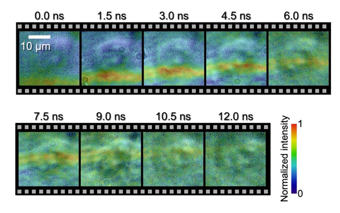 Photographs of an underwater shock wave shifting by way of a HeLa cell. Utilizing this new know-how, the researchers may see the distinction between how the shock wave moved inside and outdoors of a cell submerged in water. They famous that the outcomes urged that the cell construction shifts with the visualized wavefront place (proven within the purple/ orange line within the picture). (Picture: 2023 Saiki et al.)
The Analysis
You’re ready along with your digital camera for simply the suitable second. Abruptly, your topic speeds by and also you’ve barely clicked the shutter. Missed it. Timing could be all the things in pictures and capturing pictures at excessive pace poses a specific problem. However due to advances in digital camera know-how, as of late we are able to see the world like by no means earlier than. Whether or not it’s the sweat on a racing bicycle owner’s forehead, the main focus within the eyes of a swooping falcon or, with this newest enchancment in nanosecond pictures, the motion of a shock wave passing by way of a microscopic single cell at excessive pace. “For the primary time in historical past, so far as we all know, we’ve straight noticed the interplay between a organic cell and a shock wave, and experimentally demonstrated that the speed of the shock wave propagating contained in the cell is quicker than the surface of the cell,” defined Takao Saiki, a doctoral scholar from the Division of Precision Engineering on the College of Tokyo. “Moreover, our strategy has enabled us to exhibit high-speed pictures throughout a large time vary, which incorporates picosecond (one-trillionth of a second), nanosecond (one-billionth of a second) and millisecond (one-thousandth of a second) timescales.” Capturing clear pictures of cells with out affecting their construction or inflicting injury could be very difficult. To securely take the pictures, the researchers developed a precision optical circuit, a circuit that makes use of gentle as a substitute of electrical energy, which they named spectrum circuit. With spectrum circuit they created nondamaging laser pulses, which they set to emit at totally different timings. By combining this know-how with an current single-shot optical imaging method known as sequentially timed all-optical mapping pictures, or STAMP, they had been capable of take sequence of pictures with larger definition and fewer blur than beforehand obtainable. The workforce used the identical know-how to take a look at the results of laser ablation on glass. Laser ablation is helpful for exactly eradicating strong materials from a floor and is utilized in each trade and drugs. The researchers centered an ultrashort laser pulse simply 35 femtoseconds lengthy (one femtosecond is the same as one-quadrillionth of a second) onto a glass plate. Utilizing the spectrum circuit, they noticed the impression of the laser, the ensuing shock waves, and the impact it had on the glass over picoseconds, nanoseconds and milliseconds.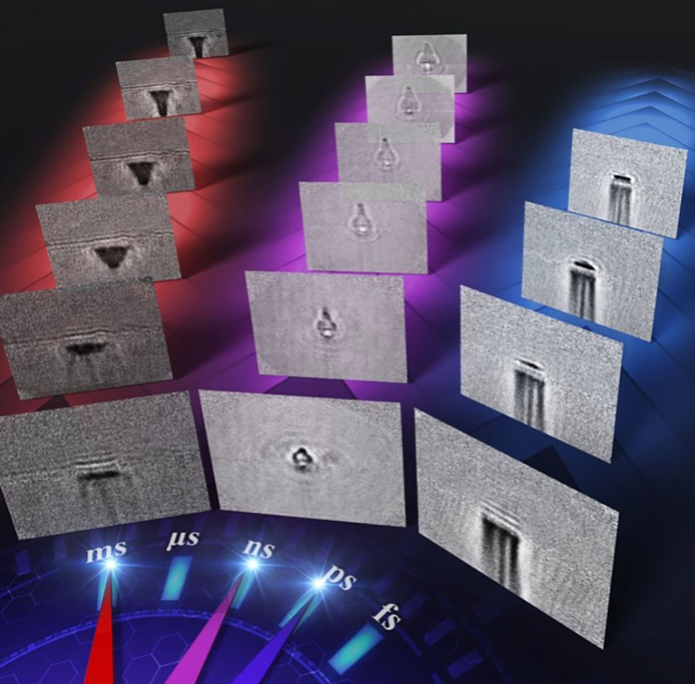 Photographs of laser ablation taken utilizing the ultrawide time vary, high-speed digital camera. Making use of this new imaging know-how, the researchers may see the propagating shock wave and plasma and the progress of laser processing over multi-timescales (about 10–100 picoseconds, about 1–10 nanoseconds, and about 1–100 milliseconds). (Picture: 2023 Saiki et al.)
“We may see the interaction between totally different bodily processes happening over time, and the way they took form,” stated Keiichi Nakagawa, an affiliate professor from the Division of Bioengineering and the Division of Precision Engineering on the College of Tokyo. “Our know-how offers alternatives to disclose helpful however unknown high-speed phenomena by enabling us to watch and analyze such ultrafast processes.
“Subsequent, we’re planning to make use of our imaging method to visualise how cells work together with acoustic waves, like these utilized in ultrasound and shock wave remedy. By doing this, we intention to know the first bodily processes that activate subsequent therapeutic results within the human physique.” The workforce additionally needs to make use of spectrum circuit to enhance laser processing methods, by figuring out the bodily parameters that might allow sooner, extra exact, extra constant and cost-effective manufacturing.
“We’ve got at all times been fascinated by the facility of visualization to know advanced phenomena. The prospect to uncover and present components of the world that had been hidden earlier than actually drew us to this area,” stated Nakagawa. “We count on to make broad contributions in numerous fields, from biomedicine to manufacturing, supplies, the surroundings and power.”
Photographs of laser ablation taken utilizing the ultrawide time vary, high-speed digital camera. Making use of this new imaging know-how, the researchers may see the propagating shock wave and plasma and the progress of laser processing over multi-timescales (about 10–100 picoseconds, about 1–10 nanoseconds, and about 1–100 milliseconds). (Picture: 2023 Saiki et al.)
“We may see the interaction between totally different bodily processes happening over time, and the way they took form,” stated Keiichi Nakagawa, an affiliate professor from the Division of Bioengineering and the Division of Precision Engineering on the College of Tokyo. “Our know-how offers alternatives to disclose helpful however unknown high-speed phenomena by enabling us to watch and analyze such ultrafast processes.
“Subsequent, we’re planning to make use of our imaging method to visualise how cells work together with acoustic waves, like these utilized in ultrasound and shock wave remedy. By doing this, we intention to know the first bodily processes that activate subsequent therapeutic results within the human physique.” The workforce additionally needs to make use of spectrum circuit to enhance laser processing methods, by figuring out the bodily parameters that might allow sooner, extra exact, extra constant and cost-effective manufacturing.
“We’ve got at all times been fascinated by the facility of visualization to know advanced phenomena. The prospect to uncover and present components of the world that had been hidden earlier than actually drew us to this area,” stated Nakagawa. “We count on to make broad contributions in numerous fields, from biomedicine to manufacturing, supplies, the surroundings and power.”
