The detailed process for making ready AFZDA NPs is illustrated in Fig. 1a. AFZ NPs had been ready by solvothermal in-situ synthesis. DOX and anti-ANG2 had been loaded through bodily adsorption in mesoporous channels of the AFZ NPs. On this work, the normalization of the vessels might improve the power of delivering oxygen and immune cells to the tumor by reconstructing the intratumoral atmosphere, as proven in Fig. 1b. AFZDA NPs might promote the shut connection of endothelial cells with perithelial cells by regulating VEGF and ANG2, normalizing the construction and performance of vessels. The hypoxia within the tumor was thus alleviated, and the infiltration of medication and anti-tumor immune cells within the tumor was promoted. The thermo-chemotherapy results had been improved via vascular normalization of the tumor, as proven in Fig. 1c.
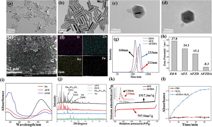
TEM imaging was used to investigate the construction of Zn0.4Fe2.6O4 NPs, Au nanorods, and AFZ, as proven in Fig. 2a, b, and c, respectively. Zn0.4Fe2.6O4 NPs with a mean measurement of ca. 6 nm had been ready with a hydrothermal methodology (Fig. 2a). The elementary composition of Zn0.4Fe2.6O4 nanoparticles was characterised by EDX, which exhibits the quantified ratio between zinc, iron, and oxygen is 0.39 to 2.61 to 0.4. The Au nanorods that had been ready with a seedless methodology had clear rod-like profiles (Fig. 2b). In Fig. 2c, Au nanorods and Zn0.4Fe2.6O4 NPs had been encapsulated in Zif-8 (AFZ). As proven in Fig. 2d, DOX loading was achieved by absorbing DOX molecules in channels and floor of the AFZ as a result of good bodily adsorption properties. SEM imaging was adopted to characterize the floor morphology and component distribution of AFZ NPs, the outcomes of that are proven in Fig. 2e–f. The AFZ NPs exhibited a slim measurement distribution and uniform distribution of O, Zn, Au, and Fe parts. The scale distribution of various samples was measured based mostly on dynamic gentle scattering (DLS) (Fig. 2g). The common measurement elevated from 160 nm (Zif-8) to 213 nm (AFZ) as a result of incorporation of Au nanorods and Zn0.4Fe2.6O4 NPs, which was in line with the outcomes of TEM imaging. The common measurement of AFZDA was 233 nm, ascribed to the encapsulation of DOX and adsorption of electronegative anti-ANG2. As well as, zeta potential values of various nanoparticles are proven in Fig. 2h. The incorporation of Zn0.4Fe2.6O4 NPs, Au nanorods, and DOX decreased the zeta potential of the Zif-8 particles. The negatively charged anti-ANG2 vastly decreased the zeta potential when it was adsorbed onto the floor of the AFZDA NPs, from 15.2 to −8.3 eV.
TEM photos of Zn0.4Fe2.6O4 NPs a, Au nanorods b, AFZ c, and AFZD d and SEM picture with component mapping e–f of AFZ; measurement distribution based mostly on DLS g and Zeta potential h of various samples; UV-vis-NIR of AFZ NPs and Au nanorods i; XRD j of Zif-8 and AFZ; The sorption and desorption curves and pore measurement distribution of AFZ and AFZDA (ok). l Time-dependent TMB consumption with AFZDA NPs
As proven in Fig. 2i, the absorption spectra of AFZD NPs, AFZ NPs, and Au nanorods of their aqueous options had been obtained by UV–vis-NIR spectroscopy. The large absorption band of AFZD NPs within the NIR area was near that typical of Au nanorods, displaying their good photothermal conversion underneath 808 nm laser irradiation.
Moreover, the XRD patterns of Au, Zn0.4Fe2.6O4 NPs, Zif-8, and AFZ NPs are proven in Fig. 2j. Zif-8 had a rhombic dodecahedron construction, whose typical diffraction peaks had been additionally discovered within the sample of AFZ NPs. Furthermore, the peaks at 38.1° and 44.3° which might be assigned to (111) and (200) planes of Au nanorods and the height at 35.8° that’s assigned to (311) aircraft of Zn0.4Fe2.6O4 NPs appeared within the sample of AFZ NPs. These outcomes point out that Zn0.4Fe2.6O4 NPs and Au nanorods had been included into the Zif-8. To evaluate the drug loading capability of AFZ, the nitrogen adsorption by AFZ and AFZDA NPs was measured by BET and the curves displayed a sort I isotherm, as proven in Fig. 2ok. The BET floor space decreased from 1517.3 m2/g to 797.54 m2/g, and the typical diameter of micropores decreased from 3.43 nm to 2.53 nm as a result of DOX loading and the adsorption of anti-ANG-2 on the floor and within the porous channels of the AFZ (Fig. 2ok). The loading capability of DOX was decided as 143 mg/g, and the loading of anti-ANG-2 was about 11 mg/g. Within the presence of H2O2, Zn0.4Fe2.6O4 nanoparticles had been progressively launched from AFZDA NPs on account of collapsing of the body construction. The colorimetric assays using 3,3′,5,5′-tetramethyl-benzidine (TMB) had been utilized to validate the peroxidase (POD)-like exercise of Zn0.4Fe2.6O4 nanoparticles. The lower of TMB focus was noticed with time (Fig. 2l).
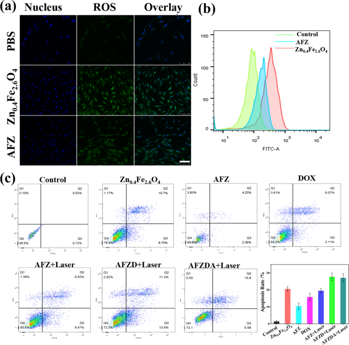
The presence of intracellular ROS was imaged by laser scanning confocal microscopy, as proven in Fig. 3a. The ROS expression dramatically elevated after therapy with Zn0.4Fe2.6O4 nanoparticles. Comparatively, the intracellular ROS was diminished when handled with AFZ nanoparticles, on account of Zif-8 coating (Fig. 3b). Annexin-v/Propidium iodide (PI) double staining package was used to judge the apoptosis of HCT116 cells with totally different therapies. The cell apoptosis was analyzed by circulation cytometry, the outcomes of that are proven in Fig. 3f. The apoptosis fee of the Zn0.4Fe2.6O4 -treated group was 20.57% on account of reactive oxygen species (ROS). Compared, the apoptosis fee of HCT116 cells handled with AFZ NPs was solely about 10%, whereas the apoptosis fee of the AFZ, AFZD, and AFZDA NPs-treated group considerably elevated underneath NIR laser irradiation (808 nm, 1.5 W/cm2, 3 min). These outcomes indicated that the Zif-8 might scale back the consequences of CDT and PTT, and the cell apoptosis induced CDT and PTT impact underneath the NIR irradiation and nanoparticles launched with the rise of temperature. The discharge of DOX from the AFZD NPs was investigated.
The temperature adjustments of the AFZD NPs and H2O with irradiation time had been recorded, as proven in Fig. 4a. At every focus, the temperature of AFZD options elevated with time underneath irradiation. The temperature improve was extra important at increased concentrations, as anticipated. On the focus of 0.5 mg/mL, the temperature of AFZD resolution elevated from 24.8 °C to 49.1 °C after the NIR laser irradiation for 600 s. In Fig. 4b, temperature elevation of the aqueous AFZD at totally different concentrations (0, 0.25, 0.5, 0.75, 1.0 mg/mL) underneath the irradiation of a NIR laser (808 nm, 1.5 W/cm2) for 600 s and shutting off the laser. As proven in Fig. 4c, τs was decided to be 167 s for the focus of 0.5 mg/mL AFZD by linear becoming cooling time (t) to detrimental pure logarithm of temperature (−ln θ). η was finally calculated to be 18.1%. These outcomes confirmed that AFZD options possessed important photothermal conversion functionality in vitro.
a Temperature variation curve of various concentrations of AFZD NPs (0.25 ~ 1.00 mg/mL) underneath the irradiation of a NIR laser (808 nm, 1.5 W/cm2) for 600 s. b Temperature elevation of the aqueous AFZD with totally different concentrations (0, 0.25, 0.5, 0.75, 1.0 mg/mL) underneath the irradiation of a NIR laser (808 nm, 1.5 W/cm2) for 600 s and shutting off the laser. c The willpower of the time fixed for warmth switch from the system utilizing linear regression of the cooling profile of the AFZD with 0.5 mg/mL focus. d Cyclic photothermal stability of AFZ NPs. e Laser scanning confocal microscopy photos of HCT116 cells cultivated with AFZ NPs. Scale bar = 50 μm. h Infrared photos of mice after administration of PBS buffer and AFZ NPs and the temperature adjustments with irradiation time (808 nm, 1.5 W/cm2). f Viability of HCT116 cells handled with the identical DOX concentrations in AFZD NPs and free DOX (n = 4, imply ± s.d. p < 0.05). g Viability of HCT116 cells handled with totally different concentrations of AFZ, AFZD and AFZDA NPs (n = 4, imply ± s.d. p < 0.05) underneath totally different circumstances. i Cumulative DOX launch from AFZD NPs (0.75 mg/mL) at totally different pH values (n = 4, imply ± s.d.)
As proven in Fig. 4d, the photothermal stability of AFZ NPs (0.75 mg/mL) was characterised. The temperature of AFZ NPs resolution elevated from 24.8 ℃ to 49.8 ℃ after NIR laser irradiation for five min. When the irradiation was off, the temperature decreased to the unique worth after 6 min. Subjected to 4 on–off cycles, the AFZ NPs didn’t present fatigue indicators, suggesting good photothermal stability.
The endocytosis of AFZ NPs was noticed by laser scanning confocal microscopy (Leica Co.). The AFZ NPs labeled with FITC had been co-incubated with HCT116 cells (the nuclei of which had been stained with DAPI) for 3 h earlier than the cells had been mounted and stained (Fig. 4e). The LSCM photos point out that nanoparticles had been internalized by the HCT116 cells, as evidenced by the overlap of inexperienced and blue (DAPI) fluorescence. Our earlier research have proven that the AFZ and AFZD NPs have glorious near-infrared absorption property. The AFZD NPs with totally different concentrations had been irradiated by a near-infrared (NIR) laser (808 nm, 1.5 W/cm2).
As proven in Fig. 4f, the CCK-8 methodology was used to review the biocompatibility and cytotoxicity, the identical DOX concentrations, AFZD NPs group has decrease cytotoxicity than free DOX, however increased cell toxicity underneath irradiated (808 nm, 1.5 W/cm2). In Fig. 4g, the viability of HCT116 cells was handled with AFZ, AFZD, and AFZDA NPs in vitro to discover the biocompatibility and therapeutic results of the nanomaterials. The cell viability of HCT 116 cells handled with the nanoparticles of various concentrations was in contrast. With rising focus of the nanoparticles, the viability of cells with totally different therapies decreased. On the identical focus, the cells handled with AFZ NPs with out laser irradiation confirmed comparatively excessive viability. When the laser irradiation was utilized for 3 min, the cell viability decreased considerably. The cells handled with AFZD and AFZDA NPs underneath laser irradiation confirmed even decrease viability for the reason that loaded DOX and/or anti-ANG-2 exert therapeutic results on the most cancers cells. On the focus of 250 μg/mL, the cell viability was decrease than 20%. These outcomes point out the good most cancers cell inhibition capacity of the nanoparticles. Additionally, the photothermal therapeutic impact underneath irradiation was confirmed.
Tumor-bearing mice had been subcutaneously injected with 2 mg/kg of AFZ NPs in addition to PBS buffer as a management, and real-time infrared imaging was carried out for the mice underneath NIR laser irradiation (808 nm, 1.5 W/cm2). The temperature of the tumor web site was detected by an infrared thermal digital camera at each 30 s. As might be seen from the temperature curves in Fig. 4h, the temperature of the tumor within the mouse injected by AFZ NPs elevated from 20.5 °C to 50.9 °C Compared, the temperature of the tumor within the mouse injected with PBS buffer solely elevated to 33.1 °C. Determine 4i exhibits the DOX launch curves of AFZD throughout a interval of 24 h at pH = 7.4 and 6.0, respectively. At pH 7.4 typical of physiological atmosphere, the cumulative launch of DOX was solely 22.6% at 24 h. It’s attribute of the endolysosomal system in most cancers cells at pH 6.0, nevertheless, greater than 90% of the loaded DOX was launched. These outcomes point out that the nanoparticles stored steady underneath physiological circumstances, which had been in a position to launch the chemo-therapeutic drug underneath intracellular circumstances.
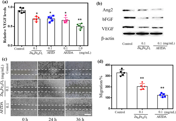
The components associated to blood vessel normalization embrace VEGF, b-FGF, and Angiopioetin. A VEGF-MV Full Package was used to tradition HUVECs, which had been then handled with Zn0.4Fe2.6O4, AFZD NPs, and AFZDA NPs for twenty-four h, respectively. As proven in Fig. 5a, the expression degree of VEGF decreased in contrast with the management group. When the HUVECs had been handled with 0.1 mg/mL AFZDA NPs, the VEGF expression decreased by 35.7%. The western blotting ends in Fig. 5b point out that the Zn0.4Fe2.6O4 NPs diminished the secretion of VEGFR1 and b-FGF, and the AFZDA NPs might inhibit the expression of Angiopioetin2.
a Zn0.4Fe2.6O4 NPs, AFZD NPs, and AFZDA NPs inhibiting VEGF of HUVECs measured by Elisa package Elisa at 24 h. b Western blot evaluation for Ang2, b-FGF, and VEGFA expression in 0.1 mg/mL Zn0.4Fe2.6O4 NPs and AFZDA NPs handled HUVECs and β-actin was used as the interior reference. c The wound-healing assay indicating the migration capacity of HUVECs utilizing Vasculife-MV full package (0 h, 24 h, and 36 h after therapy with PBS or 0.1 mg/mL Zn0.4Fe2.6O4 NPs, scale bar = 200 µm. d The migration of HUVECs after therapy 36 h. All knowledge are expressed as imply ± SD, n = 5 per group. P values had been calculated utilizing the two-paired Pupil’s t-test, handled teams vs. management group (*P < 0.05,**P < 0.01, ***P < 0.001)
Wound therapeutic experiments had been carried out to look at the consequences of AFZDA NPs on cell migration. As proven in Fig. 5c, each Zn0.4Fe2.6O4 and AFZDA NPs hampered the migration of the HUVECs to the scratch zone. The AFZDA NPs inhibited the lateral switch of the cells extra considerably, lowering the migration fee by 61% in comparison with the management group.
The results of Zn0.4Fe2.6O4 and AFZDA NPs on the vessel formation capacity of the vascular endothelial cells had been investigated. The HUVECs had been added to the 24-well plates with Matrigel and incubated in a single day. Then totally different formulations had been added to the wells, which had been then incubated for twenty-four h. Then the HUVECs had been seeded in Matrigel and cultured within the incubator for 0.5–6 h. A considerable amount of HUVECs contacted and fused to type a tube construction incubating for 1 h, which additional fashioned a community construction incubating for 3 h. Thereafter the tubular construction disintegrated progressively. The dynamic strategy of the angiopoiesis was noticed by microscopy, as proven in Fig. 6. The software program Picture J was used to investigate the variety of tubes and nodes. Within the case of therapy with 0.1 mg/mL Zn0.4Fe2.6O4 and AFZDA NPs, the variety of tubes fashioned was 18 ± 1.4 and three ± 0.8, respectively. The variety of tubes of the management V group was 31 ± 2.5 (Fig. 6b). Handled with 0.1 mg/mL AFZDA NPs, the variety of nodes fashioned was 21 ± 4.8, in comparison with that of the management V group, 114 ± 5.8 (Fig. 6c). These outcomes point out that therapy with 0.1 mg/mL AFZDA NPs for twenty-four h was able to inhibiting the tube formation of the HUVECs in vitro.
a Results of Zn0.4Fe2.6O4 NPs and AFZDA NPs on tube formation of HUVECs induced by VEGF. HUVECs had been inoculated on the Matrigel and handled with VEGF (40 ng/mL) within the presence or absence of both Zn0.4Fe2.6O4 NPs and AFZDA NPs (scale bar = 200 µm). The quantitative of tube formation b and junctions c had been evaluated after totally different therapies by angiogenesis evaluation from picture J software program. (n = 7, imply ± s.d.) *P < 0.05, **P < 0.01, ***P < 0.001 versus management
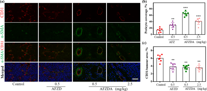
To judge the power of the AFZDA NPs to normalize the tumor vessel morphology, the maturity of the tumors within the mice handled with AFZD and AFZDA NPs was investigated. The tumor tissues had been double stained with the tumor endothelial cell marker CD31 and perithelial cell marker α-SMA. Then the tumor-bearing mice had been handled with AFZD and AFZDA NPs, respectively, each different day for 14 days. As proven in Fig. 7a, within the tumor tissues of the management group, a small quantity of α-SMA optimistic cells dispersed across the HUVECs, and the vessel construction was immature. Moreover, α-SMA optimistic cells existed within the CD31 optimistic areas. After therapy with 0.5 mg/kg AFZDA NPs, the α-SMA optimistic cells, and the CD31 optimistic cells aggregated densely and the protection of perithelial cells was 76%, as proven in Fig. 7b. The vascular construction of the tumor turned extra intact. The variety of blood vessels was diminished as compared with the management group, as proven in Fig. 7c.
a The consultant photos and morphologic evaluation of double staining of tumor sections for CD31 (endothelial cells, crimson), α-SMA (pericyte cells, inexperienced), and DAPI (nucleus, blue) after therapy with AFZD and AFZDA NPs at totally different doses ( scale bar = 50 µm); b pericyte protection after nanoparticles therapy with totally different doses and c the CD31of tumor, (n = 7, imply ± s.d.); **P < 0.01, ***P < 0.001 versus management
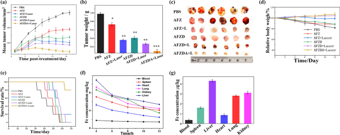
To additional verify the therapeutic results of the nanoparticles, the tumor-bearing nude mouse mannequin was constructed. The tumor-bearing mice had been injected through tail vein with 0.5 mg/kg AFZ, AFZD, or AFZDA NPs as soon as each different day. The nude mice in every group had been anesthetized and killed, and the tumors had been harvested, dissected, and noticed after 24 days of therapy. As proven in Fig. 8a–d, the therapeutic results of the nanoparticles on the tumor had been in line with the outcomes of cell experiments mentioned above. Determine 8a signifies the tumor progress inhibition impact of the nanoparticles, because the tumor progress of the mice handled with numerous formulations of nanoparticles was suppressed to totally different extents in contrast with that of the mice handled with PBS buffer. The hyperthermia impact of the AFZ NPs underneath NIR laser irradiation considerably inhibited the speedy progress of the tumor, however the subsequent tumor progress couldn’t be managed utterly. When the hyperthermia impact was mixed with DOX and anti-ANG2, the tumor progress inhibition turned extra important, as mirrored by the diminished quantity of tumor after 8 days of therapy with AFZDA NPs. The tumor weight after therapy with totally different formulations was additionally recorded. As proven in Fig. 8b, the typical tumor weight of the AFZDA NPs-treated mice group underneath laser irradiation was 0.1 g. Compared, the typical tumor weight of the AFZD NPs-treated group and that of the AFZ NPs-treated group underneath laser irradiation was 0.21 g and 0.73 g, respectively. These outcomes had been in good accordance with these of tumor quantity. Determine 8c exhibits the images of the harvested tumors from the teams with totally different therapies, which additional confirmed the therapeutic results of the nanoparticles. The burden of the group handled with AFZDA NPs-treated group underneath laser irradiation had a slight improve, which indicated that AFZDA NPs have increased safety, as proven in Fig. 8d. The survival time of tumor-bearing mice handled with totally different formulations was investigated, as proven in Fig. 8e. The mice handled with AFZDA NPs underneath irradiation had the best survival fee, and no mice died inside 60 days. The AFZDA NPs exhibited long-term anticancer exercise, and the lifetime of tumor-bearing mice was successfully prolonged. The excessive anticancer efficacy of AFZDA NPs instructed the essential function of the normalization of tumor blood vessels in tumor therapy.
a Imply tumor volumes of the mice handled with totally different formulations on the nanoparticles dose of 0.5 mg/kg (808 nm laser irradiation, 1.5 W/cm2, 3 min); b Imply tumor weights. c The {photograph} of the tumors after totally different therapies and d The burden of tumor-bearing mice. e Survival curves from numerous therapies (nanoparticles dose 0.5 mg/kg). f Imply Fe concentrations (mg/kg) in organ with time after one single injection and g three consecutive injections. (n = 5, imply ± s.d.)
To analyze the physique security of AFZDA NPs, the distribution of component Fe within the mice physique was studied. 0.5 mg/kg AFZDA NPs had been injected into the mice through tail vein, and the Fe content material in numerous organs of mice was measured, as proven in Fig. 8f. The quantity of Fe ions within the organs progressively decreased with time after injection. The AFZDA NPs primarily gathered within the liver, kidney, and lung. Then the AFZDA NPs had been consecutively injected into the mice thrice, and the Fe focus within the organs is proven in Fig. 8g. The AFZDA NPs nonetheless preferentially gathered in liver, kidney, and lung in vivo, indicating that the AFZDA NPs had been metabolized primarily in liver and kidney.
Hypoxia-inducing issue (HIF-1α) is very expressed in tumor tissues on account of hypoxia (Fig. 9). The inexperienced fluorescence (HIF-1α) was considerably diminished (AFZDA NPs group). Moreover, vascular normalization helps to alleviate hypoxia in tumor tissues via AFZDA NPs handled. The outcomes once more confirmed that the tumor hypoxia was improved due to AFZDA NPs. Reworking of tumor immune microenvironment is a brand new course of tumor remedy synergistically.
a Consultant fluorescence photos of CT26 subcutaneous tumor immunostained with CD31(endothelial cells, crimson), pimonidazole (hypoxia, inexperienced), and DAPI (nucleus, blue) staining. after therapy with AFZD and AFZDA NPs at totally different doses. b Quantification of the hypoxic space of tumor space following pimonidazole (50 mg/kg) injection and marking. (n = 5, imply ± s.d.); *P < 0.05, **P < 0.01 versus management
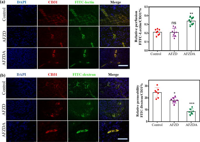
As an satisfactory blood provide is required for tumor progress, native invasion, and metastasis, tumor blood vessels play an vital function within the growth of tumors. The morphology and construction of microvessels in tumor tissues are of nice significance for understanding the therapy of tumors (Fig. 10a–b). The tumor-bearing mice had been handled with AFZ and AFZDA NPs as soon as each 2 days. FITC-lectin (inexperienced) was injected intravenously to mark the tissue vessels and CD31 (crimson) was used to label vascular endothelial cells. ANG2 in AFZDA inhibited angiogenesis and elevated vascular perfusion. It may be seen from Fig. 10a that the vascular perfusion and diameter elevated after the therapy with AFZDA NPs therapy. This indicated that the tumor blood vessels of mice handled with AFZDA NPs had important adjustments in morphology and construction, that are alerts of tumor vessel normalization. Excessive interstitial stress results in drug penetration obstacles in tumor tissues [42]. The irregular blood vessels make it troublesome for nanoparticles to diffuse into tumor tissues underneath excessive interstitial stress as a result of proliferation of tumor cells and collagen interentanglement. On this work, dextran labeled with fluorescein isothiocyanate (FITC-dextran) and CD31 had been used as vascular markers to look at the quantity of dextran infiltrating into tumor tissues from blood vessels, which represents the permeability of blood vessels. After FITC-dextran (inexperienced) was injected into the mice’s physique, part of the dextran remained within the vascular lumen and was hooked up to the floor of endothelial cells. There’s a optimistic correlation between the density of blood vessels and the quantity of dextran remaining in blood vessels in tumor tissues. The microvessel permeability might be expressed by the ratio of fluorescence depth of FITC-dextran to that of CD31. The ratio of the AFZDA NPs-treated group was decrease in comparison with the PBS group, indicating that the therapy with AFZDA NPs improved the tumor microenvironment and normalized the blood vessels, thus reducing blood permeability. The mechanism is that the gradual degradation of collagen in the course of the therapy decreases the tumor stress and normalizes blood vessels, which facilitates photothermal tumor remedy.
a The perfusion of tumor vessels measured by injection of FITC-lectin (inexperienced; crimson = CD31) after totally different therapies; b The vessel permeability noticed by labeling with FITC-dextran (scale bar = 100 µm). The permeability of tumor vessels was measured. (n = 7, imply ± s.d.); Represents no important distinction (ns), *P < 0.05, **P < 0.01, **P < 0.001 versus management
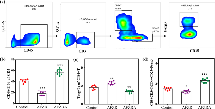
Immune T cells had been analyzed to elucidate how they performed the therapeutic function by autoimmune mechanism in the course of the therapy. Moreover, cytotoxic T cells ( CTL, CD8+ CD3+T cells) are the principle effector cells of anti-tumor immunotherapy and may actively induce tumor-specific antigens. Move cytometric evaluation is proven in Fig. 11a. The variety of CD8+T cells considerably elevated in tumor tissues of the mice handled with AFZDA NPs, accounting for 53.6% of the entire CD3+T cells (Fig. 11b). Though AFZD NPs had a superb photothermal-chemo-therapeutic impact, the expression of immune effector cells was inhibited on the tumor websites.
a Move cytometric evaluation of various teams tumors after therapy PBS, AFZD, AFZDA underneath laser (808 nm, 1.5 W/cm2, 3 min). Quantification of the info from the experiment and the proportion of CD8+T of CD3+T cells b, the proportion of Treg of CD4+T cells c, the proportion of CD8+ CD3+T of CD4+ CD25+Foxp3 cells (CTL/ Treg) d. (n = 7, imply ± s.d. *P < 0.05, **P < 0.01, **P < 0.001 versus management)
Tregs can inhibit the immune response as a subset of CD4+T cells. As proven in Fig. 11c, the expression degree of Treg was 19.24%, 21.68%, and 16.93% in tumors handled with PBS, AFZD NPs, and AFZDA NPs, respectively. These outcomes confirmed that the differentiation of immunosuppressive cells was successfully inhibited by reversing the tumor hypoxia. The AFZDA NPs improved the tumor infiltration of CD8+T cells via blood vessel normalization and diminished the differentiation of the Tregs (CD4+CD25+Foxp3) to some extent. Thus, the ratio of CD8+T cells to Tregs elevated, as proven in Fig. 11d.
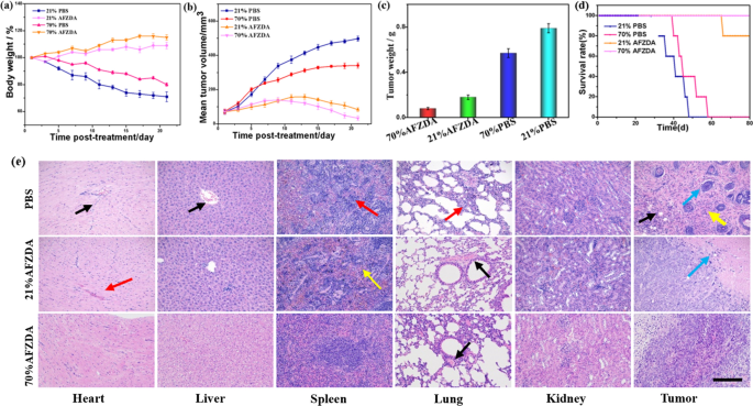
The oxygen focus was additional studied throughout tumor photothermal/ chemo-therapy therapy. After 21 days of therapy, the mice had been anesthetized and killed, and the antitumor results of various therapy teams had been measured. The physique weight, tumor weight, and tumor quantity of mice in every group had been in contrast, as proven in Fig. 12a–c. It was discovered that hyperoxia might be used as a therapy to inhibit tumor progress and decelerate the tumor progress fee in mice. The physique weight of the management group decreased by practically 30% in 21 days, whereas solely the high-oxygen therapy group decreased by about 20%. No obvious weight reduction was noticed within the respiratory 70% oxygen AFZDA therapy group(Fig. 12a). The tumor quantity and weight, in contrast with the management group, weren’t solely rapidly inhibited, however the authentic tumor was shrinking. The ultimate common tumor weight was lower than 0.1 g (Fig. 12c), indicating that the AFZDA hyperoxia group had an apparent anti-tumor impact. The survival time of mice in every group handled by totally different strategies was analyzed experimentally as proven in Fig. 12d. No dying of mice occurred inside 80 days within the AFZDA hyperoxia group. AFZDA NPs confirmed long-term anticancer exercise. It was proved that the hyperoxia clearly inhibited most cancers mixed with photothermal/chemo-therapy and successfully prolonged the survival time of tumor-bearing mice.
a Imply weight of tumor-bearing mice from respiratory totally different concentrations of oxygen after therapy PBS, AFZDA. b Imply tumor quantity of various teams; c imply tumor weights (n = 4, imply ± s.d. P < 0.05) and d survival curves (AFZDA NPs dose 0.5 mg/kg) (n = 5). e Hematoxylin–eosin stained photos of tissues and organs obtained. Scale = 200 μm
The organs of mice and tumor tissues had been stained by H&E, as proven in Fig. 12e. The toxicity was additional studied throughout therapy in vivo. In contrast with the management group, the therapy group of tissues and organs, no apparent irritation and necrosis occurred. The construction of the myocardium was regular, and no apparent degeneration was discovered. There may be hyperemia within the stroma, as proven by the crimson arrow. Within the PBS group, apparent inflammatory cells had been filtrated within the tissue, as proven within the black arrow. A small variety of hepatocytes within the management group confirmed slight edema (yellow arrows). The general construction of the spleen was regular, with connective tissue hyperplasia (yellow arrow). Numerous granulocyte infiltrates had been seen within the management group (crimson arrows). No alveolar fusion and growth, however the management group confirmed important thickening (crimson arrow). The epithelial cell construction was full within the renal tissue and the glomerular construction was seen (as proven within the black arrow). The blue arrow confirmed tumor cells. Numerous inflammatory cells had been noticed within the PBS and AFZD teams (yellow arrows). The black arrows point out that the inflammatory cells infiltrated the tumor tissue. The outcomes of H&E staining confirmed that AFZDA NPs had no unwanted side effects on animals throughout therapy.
CD8+T cells are one of many foremost effector cells of anti-tumor immunotherapy and may actively induce tumor-specific antigens. The tumor-burdened Balb/c mice respectively had been handled at 21% and 70% oxygen concentrations for 4 h/day for 4 days underneath NIR laser irradiation (808 nm, 1.5 W/cm2, 3 min). Move cytometric evaluation was proven in Fig. 13a. The proportion of CD8+T cells of spleen tissue was 18.3% in clean group mice, whereas 19.9% in AFZDA therapy group underneath 21% oxygen focus. The proportion of CD8+T cells within the group handled with 70% oxygen was increased than that within the 21% oxygen group (22% and 21%, respectively). The outcomes have proven that prime oxygen focus contributes to the differentiation of CD8+T cells in vivo. Tregs can inhibit the immune response as a subset of CD4+T cells. As proven in Fig. 13b, the expression degree of Treg was 7.04% and 5.68% in tumors handled with PBS and AFZDA teams, respectively. When underneath 70% Oxygen, Treg expression was down-regulated to 4.8% within the PBS group and three.65% in AFZDA group, respectively. The end result confirmed that it successfully inhibited the differentiation of immunosuppressive cells by respiratory hyperoxia.
Move cytometric evaluation of the spleen from totally different therapy mice, the proportion of CD8+T cells of CD3+T cells a and Treg of CD4+T cells b (n = 5, imply ± s.d.). c The partial stress of oxygen in tumors with the depth of the tumor, (n = 5, imply ± s.d.). The fluorescence imaging of HIF-1α d and CD73 e in vivo. Scale bar = 200 μm; quantification of HIF-1α and CD73 in tumors. (n = 7, imply ± s.d.); **P < 0.01, *P < 0.05 versus management
Respiratory a excessive focus of oxygen improves oxygen partial stress (Fig. 13c). Moreover, the hypoxia-inducing issue (HIF-1α) is very expressed in tumor tissues on account of hypoxia (Fig. 13d). After 70% oxygen therapy, the inexperienced fluorescence (HIF-1α) was considerably diminished. Moreover, vascular normalization helps to alleviate hypoxia in tumor tissues via AFZDA NPs handled. As everyone knows, the cells are metabolized primarily by glycolysis, resulting in immunosuppression within the absence of oxygen. Hyperoxia remedy can inhibit tumor hypoxia and scale back intracellular adenosine formation. The expression of CD73 (in Fig. 13e) was blocked by the change in metabolism. The outcomes once more confirmed that the tumor hypoxia was improved due to AFZDA NPs. Reworking of tumor immune microenvironment is a brand new course of tumor remedy synergistically.
When it comes to immune cells, the AFZDA + 70percentO2 group confirmed important variations with AFZDA therapy. In the meantime, when it comes to cytokines, the experiment additional characterised the adjustments in tumor immune microenvironment throughout AFZDA therapy with a excessive focus of oxygen. In contrast with AFZDA, AFZDA + 70percentO2 couldn’t solely scale back anti-inflammatory cytokines (TGF-β and IL-10) extra successfully but in addition improve the extent of anti-tumor pro-inflammatory cytokines IFN-γ in tumors (Fig. 14). The mixture of the AFZDA and high- focus oxygen had the strongest anti-tumor immune enhancement.
a TGF-β immunohistochemistry of tumor tissue, Scale = 200 μm. b Quantification of TGF-β in tumors. c TGF-β, d IFN-γ, and c IL-10 contents in tumors had been examined utilizing circulation cytometry. (n = 5, imply ± s.d.); represents no important distinction (ns); **P < 0.01, ***P < 0.001, versus management
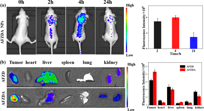
To check the distribution and metabolism of the AFZDA NPs within the mice physique, the mice had been administered with the nanoparticles and had been fluorescently imaged in vivo. As proven in Fig. 15a, the AFZDA NPs had been distributed in numerous elements of the mouse physique at 2 h after injection, and plenty of nanoparticles gathered within the tumor, as mirrored by the excessive sign depth on the tumor web site. At 4 h after injection, the distribution of the AFZDA NPs within the mouse physique was extra apparent, and nonetheless extra nanoparticles gathered within the tumor. At 24 h after injection, the AFZDA NPs within the mouse physique dramatically decreased as a result of metabolism of the nanoparticles. The fluorescence sign on the tumor web site, nevertheless, was diminished solely barely, suggesting the excessive retention of the nanoparticles within the tumor.
a In vivo fluorescence imaging of the CT26 tumor-bearing mice at 2, 4, and 24 h after intravenous injection of AFZDA NPs (left). The quantitative evaluation of fluorescent alerts from the tumors at 2 h, 4 h, and 24 h (n = 3, imply ± s.d.) (proper). b Fluorescence imaging of the tumor, coronary heart, liver, spleen, lung, kidney at 24 h after the mice had been handled with AFZD NPs and AFZDA NPs, respectively. The quantitative evaluation of fluorescence alerts for the organs at 24 h publish tail vein injection (n = 3, imply ± s.d.)
Then the tumor and main organs had been harvested from the mouse at 24 h after injection and had been imaged fluorescently. These harvested from a mouse handled with AFZD NPs underneath the identical circumstances had been additionally imaged for comparability. As proven in Fig. 15b, the little sign was detected from the center, spleen, and lung of each teams. The tumor, liver, and kidney emitted fluorescence, indicating that the nanoparticles remained in them. In contrast with the AFZD NPs, extra AFZDA NPs remained within the tumor, suggesting the upper retention of the AFZDA NPs that favored the tumor remedy.
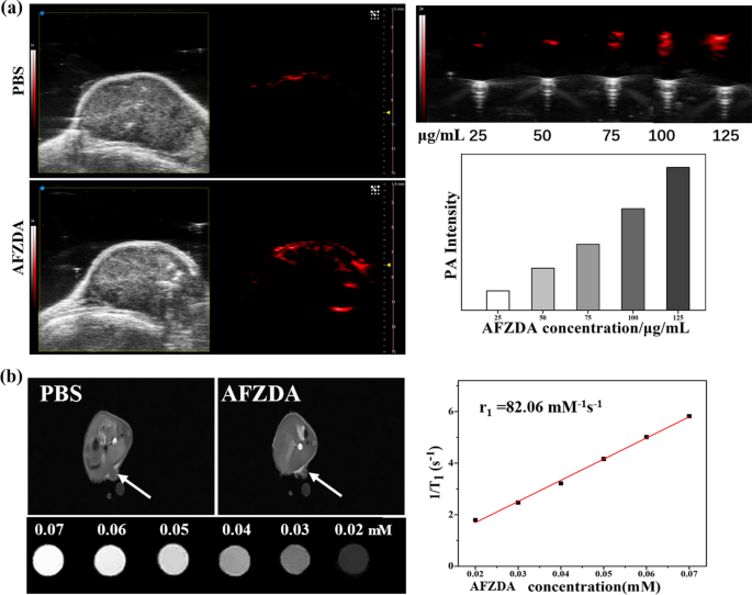
PAI was used for monitoring the tumor microenvironment in vivo. As is proven in Fig. 16a, the sign depth elevated with rising focus of AFZDA NPs in vitro. Moreover, the PA photos of the AFZDA NPs-treated group confirmed robust structural data on the tumor web site in vivo. Whereas multimodal imaging can help within the prognosis and therapy of tumors, PAI allows real-time imaging to facilitate the analysis of therapeutic results.
a The PA photos of PBS and AFZDA NPs, and the quantitative PA alerts of AFZDA NPs with concentrations had been analyzed. b The MR photos of PBS and AFZDA NPs in vivo and T1-weighted magnetic resonance imaging of AFZDA NPs with concentrations in vitro, and T1 rest fee (r1) of AFZDA NPs had been measured
The Zn0.4Fe2.6O4 NPs had been used as an MRI distinction agent each in vitro and in vivo. At 30 min after injection of 100 μL of PBS or AFZDA NPs (0.05 mM), the tumor web site of the AFZDA-treated group might be extra clearly distinguished. In the meantime, the MR alerts had been linearly enhanced because the AFZDA NPs focus elevated in vitro, as proven in Fig. 16b. The relief fee (r1) of AFZDA NPs was as excessive as 82.06 mM−1 s−1.


