Physicochemical characterization of MLNPs
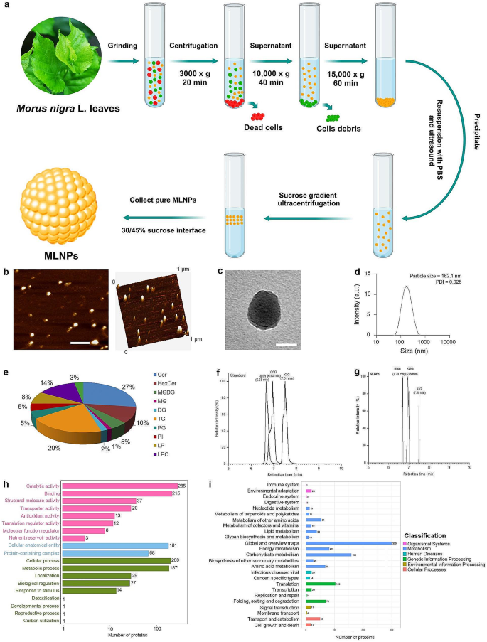
MLNPs have been extracted from recent Morus nigra L. leaf juice and purified via differential centrifugation and sucrose density gradient ultracentrifugation (Fig. 1a). Pushed by the sucrose gradients, MLNPs have been primarily distributed within the sucrose density layer of 30/45%. Atomic pressure microscopy (AFM) and transmission electron microscopy (TEM) revealed that MLNPs appeared as exosome-like spherical particles with a diameter of roughly 100 nm following dehydration (Fig. 1b, c). The additional evaluation of dynamic gentle scattering (DLS) unveiled that MLNPs possessed a hydrodynamic particle dimension of 162.1 nm and exhibited an exquisitely uniform dimension distribution (polydispersity index; PDI = 0.025), as proven in Fig. 1d. The imply diameters of MLNPs obtained by AFM and TEM have been considerably smaller than that decided by DLS, most likely attributing to their shrinkage earlier than AFM/TEM and swell throughout DLS assessments.
Physicochemical characterizations of MLNPs. (a) Extraction of MLNPs from Morus nigra L leaves. (b) AFM photos (scale bar = 200 nm), (c) TEM photos (scale bar = 50 nm), (d) hydrodynamic particle dimension distribution, and (e) lipid compositions of MLNPs. Chromatograms of (f) commonplace and (g) MLNPs. (h) GO secondary classification statistical chart and (i) KEGG annotated statistical chart of MLNPs
The lipidomic evaluation revealed that MLNPs have been predominantly comprised of ceramides (Cer, 27%), triglycerides (TG, 20%), hemolytic phosphatidylcholine (LPC, 14%), and hexose ceramides (HexCer, 10%). These constituents considerably contributed to the steadiness of MLNPs whereas orchestrating the regulation of numerous physiological processes, encompassing macrophage activation, tissue reworking, and wound therapeutic. We discovered that hemolytic phospholipids have been current in MLNPs, which might facilitate lipid fusion and improve the switch of vesicular contents to cells [27]. In the meantime, MLNPs have been discovered to include 5.0% of monogalactodiglycerol (MGDG) (Fig. 1e), which exhibited a selected affinity in the direction of the asialoglycoprotein receptor (ASGPR) over-expressed on the floor of liver tumor cells, thereby facilitating environment friendly reside tumor-targeted supply of bioactive molecules [28]. As reported, mulberry leaves harbored a plethora of small energetic molecules, encompassing an in depth vary of flavonoids, polyphenols, polysaccharides, alkaloids, and different small compounds. Flavonoids in nanovesicles have been decided utilizing an ultra-performance liquid chromatography system and comparability with the mulberry metabolomics database (MMHub) [29, 30]. It was detected that MLNPs contained 3 major flavonoids: rutin, quercetin 3-O-glucoside (Q3G), and kaempferol-3-O-glucoside (K3G) (Fig. 1f, g). The Gene Ontology (GO) purposeful database annotated a complete of 1,312 genes for complete purposeful classification, of which 602 genes take part in numerous molecular features distributed throughout 8 distinct classes, notably inclusive of catalytic exercise and binding. Moreover, 561 genes have been recognized to be concerned within the biochemical processes, which have been additional divided into 9 seed features, with mobile and metabolic processes being the first features. Ultimately, 249 genes have been discovered to be related to mobile elements consisting solely of mobile anatomical entities and protein-containing complexes (Fig. 1h).
KEGG pathway evaluation revealed that 34 proteins in MLNPs have been related to human ailments and liver cancer-associated signaling pathways (Fig. 1i). The expansion components (TGF-α, TGF-β, IGF-II, and HGF) and Wnt signaling pathway are reactivated throughout tissue regeneration, cell renewal, and sure pathological circumstances (e.g., premalignant illness and most cancers). The Wnt/β-catenin protein pathway was predominantly quiescent within the mature and wholesome liver [31]. A big proportion of liver tumors harbored mutations in genes encoding the essential elements of the Wnt/β-catenin protein signaling pathway. These findings present an perception into the molecular mechanism by which mulberry nanovesicles inhibit hepatoma cells.
In vitro anti‑tumor actions of MLNPs
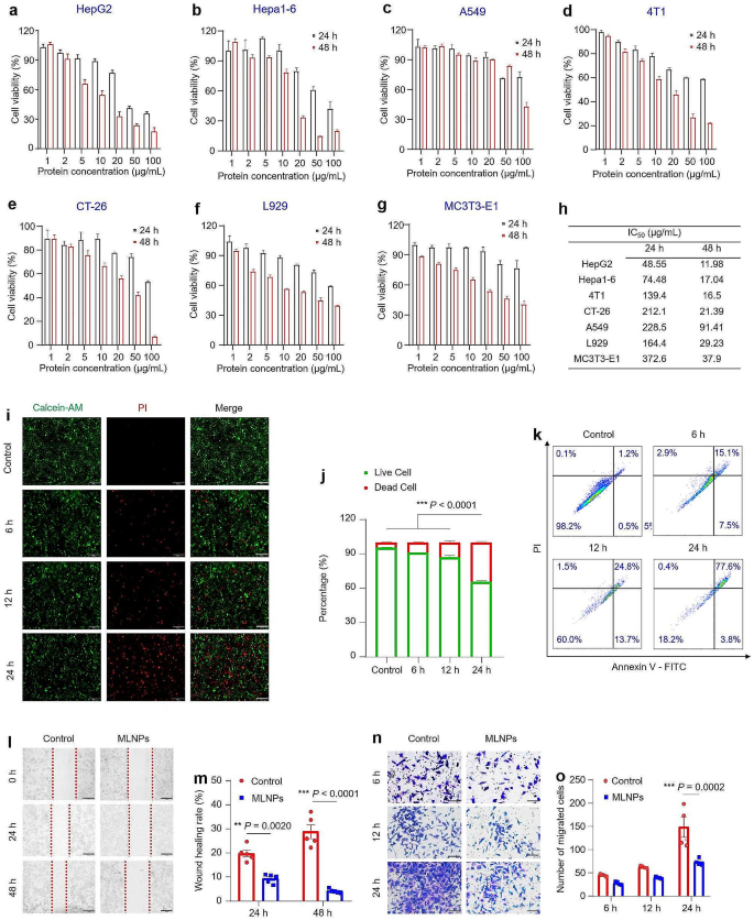
MTT assay was used to judge in vitro inhibitory potential of MLNPs towards numerous tumor and wholesome cell strains. The dose-dependent and time-dependent anti-proliferative results of MLNPs have been noticed throughout all 6 cell strains (Fig. 2a-g). Amongst HepG2, Hepa1-6, CT-26, 4T1, and A549 cells, MLNPs exhibited most cytotoxicity in the direction of HepG2 and Hepa1-6 cells, and IC50 values of the MLNP-treated HepG2 and Hepa1-6 cells have been 48.6 and 74.5 μg/mL after a 24-hour publicity, and 12.0 and 17.0 μg/mL after 48 h, respectively (Fig. 2g). The strongest anti-proliferative impact on HepG2 and Hepa1-6 cells is likely to be ascribed to the presence of galactose finish group-contained glycolipids within the MLNPs that particularly goal hepatoma cells. These outcomes reveal that MLNPs have a promising potential for HCC remedy. Notably, MLNPs demonstrated comparatively decrease toxicities towards wholesome cells than hepatoma cells (Fig. 2f, g), suggesting their good biocompatibility.
In vitro anti‑tumor actions of MLNPs. MTT assay was used to evaluate the potential toxicity of MLNPs towards (a) HepG2, (b) Hepa1-6, (c) A549, (d) 4T1, (e) CT26, (f) L929, and (g) MC3T3-E1 cells after co-incubation for twenty-four and 48 h, respectively. Every level represents the imply ± S.E.M. (n = 5). (h) The IC50 values of MLNPs towards totally different cell strains. Every level represents the imply ± S.E.M. (n = 5). (i) Fluorescence photos of Hepa1-6 cells stained with Calcein-AM/PI. Stay cells have been stained inexperienced with calcein-AM, and lifeless cells have been stained purple with PI (Scale bar = 200 μm). (j) Quantitative evaluation of the fluorescence depth of reside or lifeless cells. Every level represents the imply ± S.E.M. (n = 5; *p < 0.05, **p < 0.01, and ***p < 0.001). (ok) Apoptosis impact of Hepa1-6 cells with the remedy of MLNPs for six, 12, and 24 h, respectively. (l) Migration of Hepa1-6 cells with or with out the remedy of MLNPs for twenty-four and 48 h, respectively (Scale bar = 200 μm). (m) Wound therapeutic charges of Hepa1-6 cells within the presence or absence of MLNPs utilizing ImageJ software program. Every level represents the imply ± S.E.M. (n = 5; *p < 0.05, **p < 0.01, and ***p < 0.001). (n) The transwell migration capability of Hepa1-6 cells with the remedy of MLNPs for six, 12, and 24 h, respectively (Scale bar = 100 μm). (o) Cell counts of transwell migration at 6, 12, and 24 h utilizing ImageJ software program. Every level represents the imply ± S.E.M. (n = 4; *p < 0.05, **p < 0.01, and ***p < 0.001)
Subsequent, we used reside/lifeless cell double staining to confirm the toxicity of MLNPs to hepatoma cells. Calcein-AM enters dwelling cells and emits inexperienced fluorescence after being damaged down by esterases, whereas PI solely enters lifeless cells and emits purple fluorescence when binds to DNA [32]. As depicted in Fig. 2i, Hepa1-6 cells demonstrated a discernible time-dependent augmentation in purple fluorescence intensities subsequent to co-incubation with MLNPs for six, 12, and 24 h, in distinction to the management group. The quantitative outcomes revealed a survival price of roughly 60% of cells following a 24-hour remedy with MLNPs (Fig. 2j), a discovering in concordance with the outcomes obtained from the MTT assay. Furthermore, Annexin V-FITC/PI staining revealed that superior apoptosis or necrosis occurred in 77.6% of Hepa1-6 cells receiving the remedy of MLNPs at 24 h (Fig. 2ok). Tumor cell metastasis includes migration and invasion, which causes over 90% most cancers deaths [33, 34]. To guage the impacts of MLNPs on tumor invasion and metastasis, we carried out cell scratch and migration assays. It was noticed that MLNPs considerably decreased wound therapeutic charges to 9.3% and 4.1% after co-incubation for twenty-four and 48 h, respectively (Fig. 2l, m), indicating their substantial capability to inhibit the migration of hepatoma cells. In alignment with the observations from the cell scratch assay, it was noteworthy that MLNPs confirmed a time-dependent affect in impeding the invasion and metastasis of tumor cells inside an outlined temporal vary (Fig. 2n, o). Subsequently, we posit that MLNPs could possibly be exploited as a promising pure nanomedicine for combating metastatic liver cancers.
In vitro mobile uptake and anti‑tumor mechanism of MLNPs
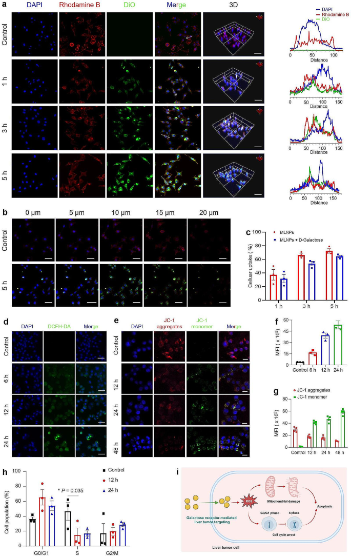
The power of nanomedicines to be internalized by focused cells is an important issue that impacts its efficacy [35]. Thus, we assessed the mobile uptake profiles of MLNPs by hepatoma cells. Since MLNPs lacked fluorescence however possessed lipophilic properties, they have been labeled with a lipophilic dye (DiO). It was detected that inexperienced fluorescence alerts inside Hepa1-6 cells progressively elevated with their elongated incubation with MLNPs, which overlapped with purple fluorescence of the cytoskeleton (Fig. 3a-c). This remark substantiates the efficient internalization of MLNPs by Hepa1-6 cells, with predominant distribution noticed inside the cytoplasm. Following co-incubation durations of 1, 3, and 5 h with MLNPs, Hepa1-6 cells exhibited uptake percentages of 37.3%, 66.0%, and 72.5%, respectively (Fig. 3c). In distinction, L929 cells displayed notably decrease uptake percentages, measuring 4.7%, 20.0%, and 30.8%, respectively (Fig. S1). Moreover, our investigation revealed decrements within the uptake percentages of MLNPs by Hepa1-6 cells following co-culture with free galactose, with reductions of 5.8%, 12.7%, and eight.1% noticed after 1, 3, and 5 h, respectively. This underscores the strong liver tumor-targeting capability of MLNPs via the galactose receptor-mediated method.
In vitro mobile uptake and antitumor mechanism of MLNPs. (a) CLSM photos and fluorescence distribution profiles (Scale bar = 50 μm). (b) CLSM cross-section photos of 5-layered mobile uptake of DiO-labeled MLNPs (inexperienced) after incubation for five h. Hepa1-6 cells have been labeled with DAPI (blue) and Rhodamine phalloidin (purple) (Scale bar = 50 μm). (c) Percentages of DiO-labeled MLNPs internalized by Hepa1-6 cells for 1, 3, and 5 h, respectively. Every level represents the imply ± S.E.M. (n = 3). (d) CLSM photos and (f) quantification of ROS adjustments in Hepa1-6 cells labeled with Hoechst 33,342 (blue) after co-incubation with MLNPs for six, 12, and 24 h, respectively (Scale bar = 50 μm). Every level represents the imply ± S.E.M. (n = 3). (e) CLSM photos and (g) quantification of JC-1 and Hoechst 33,342 stained Hepa1-6 cells after co-incubation with MLNPs for 12, 24, and 48 h, respectively (Scale bar = 20 μm). Every level represents the imply ± S.E.M. (n = 4). (h) Cell cycle evaluation of Hepa1-6 cells after co-incubation with MLNPs for 12 or 24 h by FCM. Every level represents the imply ± S.E.M. (n = 3; *p < 0.05). (i) Schematic illustration of the pro-apoptotic mechanism of MLNPs towards liver tumor cells
Intracellular ROS can induce oxidative injury to DNA, proteins, and lipids, finally resulting in the apoptosis of tumor cells [36]. We thus investigated the ROS ranges in Hepa1-6 cells receiving the remedy of MLNPs. It was detected that the management cells (with out MLNP remedy) confirmed negligible inexperienced fluorescence alerts. Quite the opposite, a gradual augmentation within the inexperienced fluorescence depth was noticed inside Hepa1-6 cells receiving the remedy of MLNPs for various durations (6, 12, and 24 h), reflecting that MLNPs might successfully induce ROS era in hepatoma cells (Fig. 3d, f). Lack of mitochondrial membrane potential is a trademark occasion throughout apoptosis [37]. JC-1 probe has been generally utilized to detect the variations of mitochondrial membrane potential. In precept, mitochondrial membrane potential will lower in apoptotic cells, and JC-1 molecules can’t be enriched of their mitochondrial matrix and exist as monomers that excite inexperienced fluorescence below the FITC channel of confocal microscope [38]. As seen in Fig. 3e, g, a discernible attenuation in purple fluorescence and a concurrent augmentation in inexperienced fluorescence have been noticed following the administration of MLNPs. These findings signify the capability of those naturally derived MLNPs to induce structural injury to the mitochondrial membrane. Moreover, PI staining was carried out to discover the capability of MLNPs in inducing cell cycle arrest. Determine 3h illustrated that after the remedy with MLNPs for 12 and 24 h, there was a notable elevation within the share of cells within the G0/G1 section, concomitant with a decrement within the proportion of cells within the S section. These observations reveal that MLNPs can block the cell cycle of Hepa1-6 cells and cause them to stagnate within the G0/G1 section (Fig. 3i).
In vivo biosafety analysis of MLNPs
Biosafety is a essential issue for the medical translation of nanomedicines [39]. We thus evaluated the in vivo biosafety of MLNPs through intravenous injection and oral administration. It was discovered that MLNPs exhibited wonderful hemocompatibility in contrast with the optimistic management group, as evidenced by the absence of blood circulation issues or vital hemolysis at any dosages (Fig. S2a, b). The physique weight of mice in every group remained secure with none vital decline (Fig. S3a). As depicted in Fig. S3b-h, oral administration of MLNPs demonstrated no observable deviations in organ indices (particularly liver and kidney) and biochemical markers when in comparison with the wholesome group. Conversely, the intravenous administration of MLNPs resulted in a notable improve in AST ranges and a concurrent discount in TBIL and GGT ranges (AST, TBIL, and GGT being pivotal markers for assessing liver perform). These findings indicate a possible hepatic toxicity related to intravenous administration of MLNPs, highlighting the relative biosafety of oral MLNPs. ALT and AST are current within the coronary heart, which is able to enter the blood stream from the broken coronary heart tissues. These two parameters didn’t considerably improve after oral administration compared with the management group (Fig. S3c, d). When it comes to renal perform, we discovered that creatinine (CRE) and blood urea nitrogen (BUN), which replicate kidney perform, confirmed no apparent adjustments amongst these three teams (Fig. S3g, h). These outcomes reveal that oral MLNPs exert negligible toxicity to the guts and kidney. As well as, the blood parameters in all teams have been inside the regular ranges, indicating that MLNPs didn’t trigger hematological toxicity (Fig. S4). The tissue morphology of the 5 precept organs from the remedy teams exhibited no vital deviation from that of the wholesome management group (Fig. S5), affirming the distinctive biosafety profile of MLNPs after oral administration.
In vivo bio‑distribution and liver focusing on of MLNPs
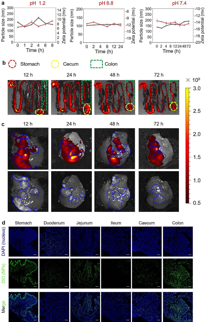
The oral route is likely one of the most most popular approaches for drug supply as a result of its comfort, cost-effectiveness, and excessive affected person compliance [40]. The steadiness of therapeutics within the GIT is an important prerequisite for oral administration [41]. Thus, we investigated the steadiness of MLNPs within the simulated gastric, small intestinal, and colonic fluids. Determine 4a revealed that MLNPs possessed comparatively secure particle sizes and floor prices in the course of the incubation in numerous simulated options, suggesting their stability within the GIT. The strong preservation of the structural integrity of MLNPs ensured their unblemished journey from the GIT to the liver tumors. DiR is a typical near-infrared fluorescent probe, which might bind to the phospholipid bilayer construction of LNPs. Consequently, we employed DiR as a labeling agent for MLNPs to trace their biodistribution inside HCC mouse mannequin. It was noticed that after oral administration, DiR-labeled MLNPs maintained within the GIT over 72 h (Fig. 4b). Strikingly, the fluorescence alerts of MLNPs have been completely overlapped with the liver tumors (Fig. 4c), demonstrating their intrinsic liver tumor-targeting capability.
In vivo biodistribution and liver focusing on profiles of MLNPs. (a) Stabilities of MLNPs within the simulated gastric, small intestinal, and colonic fluids. (b) Ex vivo fluorescence photos of the GIT within the HCC mouse mannequin receiving oral administration of DiR-MLNPs at totally different time factors (12, 24, 48, and 72 h). (c) Ex vivo tumor-targeted fluorescence photos and bio-distribution of DiR-labeled MLNPs within the orthotopic liver most cancers mouse mannequin. (d) Fluorescence photos of the GIT sections from mice receiving oral administration of DiO-MLNPs (Scale bar = 100 μm). Every level represents the imply ± S.E.M. (n = 3)
The fluorescence depth emanating from DiR-labeled MLNPs reached its zenith inside the GIT following 24 h of oral administration, as depicted in Fig. 4b. Subsequently, a detection was carried out to establish the particular absorption website inside the GIT at this designated time level. Determine 4d confirmed that apparent inexperienced fluorescence alerts (MLNPs) have been detected within the abdomen, jejunum, and colon. Furthermore, these inexperienced alerts have been enriched within the epithelial layer of the abdomen, whereas they have been primarily current within the mucosa of the jejunum and colon. These observations recommend that MLNPs won’t be absorbed within the abdomen however within the jejunum and colon, the place they enter the circulatory system and accumulate within the liver tumors through galactose receptor-mediated focusing on.
In vivo therapeutic outcomes of MLNPs towards HCC
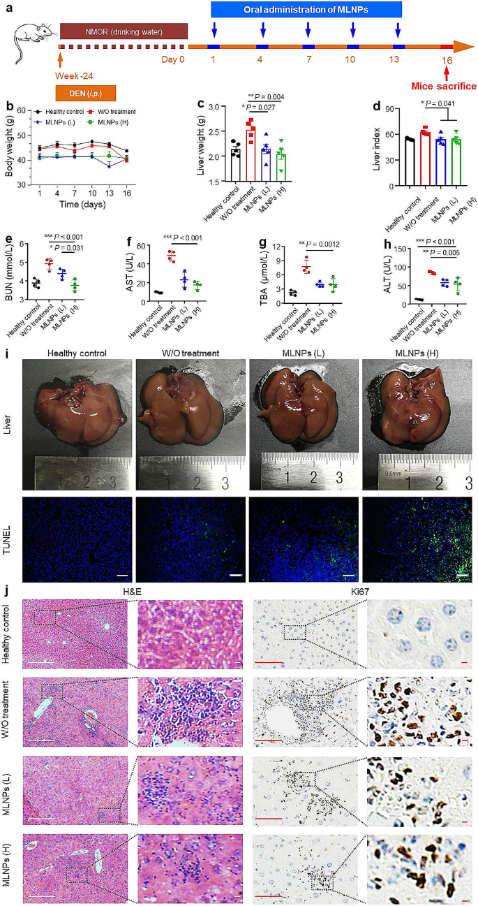
The anti-liver tumor impact of MLNPs was assessed on the premise of the DEN/NMOR-induced orthotopic liver most cancers mouse mannequin, which might carefully mimic human liver most cancers. Following the institution of an orthotopic HCC mannequin, mice have been subjected to oral administration of MLNPs on days 1, 4, 7, 10, and 13 (Fig. 5a). No obvious disparity in physique weight was discovered amongst all mouse teams throughout all the investigations (Fig. 5b). We additional discovered that in contrast with the wholesome management group, the management group (with out therapies) possessed elevated liver weight (Fig. 5c) and liver index (Fig. 5d). Nevertheless, oral administration of MLNPs (low dose: 2.5 mg/kg; excessive dose: 5 mg/kg) led to vital reductions in liver weight and liver index. Furthermore, treating MLNPs considerably improved the hepatic features of mice with orthotopic liver tumors. Particularly, the group handled with MLNPs (L) demonstrated reductions of 1.1-, 2.1-, 2.0-, and 1.5-fold within the ranges of BUN (Fig. 5e), AST (Fig. 5f), TBA (Fig. 5g), and ALT (Fig. 5h), respectively. As well as, we discovered that rising the remedy quantities of MLNPs barely decreased the liver perform indices.
In vivo therapeutic outcomes of MLNPs towards HCC. (a) Schematic diagram of the institution means of orthotopic liver most cancers mouse mannequin and remedy course of. (b) The physique weight, (c) liver weight, and (d) organ index of orthotopic liver most cancers mice with the remedy of MLNPs. Every level represents the imply ± S.E.M. (n = 5; *p < 0.05 and **p < 0.01). The quantities of (e) BUN, (f) AST, (g) TBA, and (h) ALT within the serum. Every level represents the imply ± S.E.M. (n = 4; *p < 0.05, **p < 0.01, and ***p < 0.001). (i) Consultant digital photographs, TUNEL (Scale bar = 100 μm), (j) H&E (Scale bar = 100 μm), and Ki67 (Scale bar = 100 μm) staining photos of the liver tissues from mice receiving oral remedy of MLNPs. Every level represents the imply ± S.E.M. (n = 3)
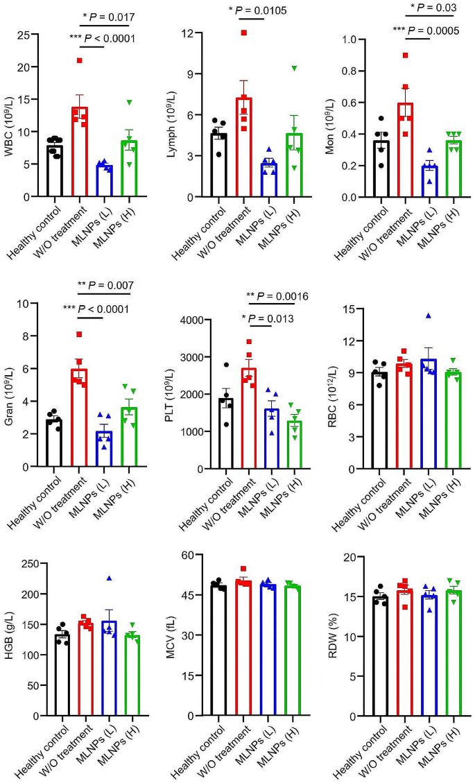
Subsequent, we decided the morphologies of livers from numerous mouse teams. Determine 5i illustrated that the teams handled with MLNPs (L and H) exhibited markedly ameliorated hepatic histology, contrasting with the liver morphology noticed within the untreated management group. We additionally discovered that the indices (Fig. S6a) and tissue morphologies (Fig. S6b) of different organs (coronary heart, spleen, lung, and kidney) from the MLNP (L and H)-treated teams had no vital variations, in contrast with these from the wholesome management group (Fig. S6a). The H&E staining photos revealed the presence of early-stage major liver tumor within the management group (with out remedy), as evidenced by hepatocyte nuclear division and vacuolar deformation. Nonetheless, oral MLNPs (L and H) considerably decreased the presentation of major liver tumors (Fig. 5j). The mix of TUNEL and Ki67 assays has been employed to label the apoptotic and proliferative profiles of cells within the tumor tissues, respectively. Remarkably, the livers from the MLNP (H)-treated group displayed probably the most intense TUNEL inexperienced fluorescence and the bottom Ki67 positivity (Fig. 5i, j). The hematological evaluation revealed that the management group, devoid of any remedy, manifested heightened counts of white blood cells, encompassing lymphocytes, neutrophils, and monocytes. These counts surpassed these noticed within the wholesome management group, indicative of inflammatory responses provoked by liver tumors (Fig. 6). Curiously, following remedy with low/excessive doses of MLNPs, the degrees of varied leukocytes exhibited vital reductions and approached normalcy. These findings recommend that oral remedy of MLNPs can effectively retard the development of liver tumors.
Impacts of oral MLNPs on intestinal microbiota
The intestine microbiome has just lately been acknowledged as a significant environmental issue within the pathobiology of many ailments. All through the previous a long time, quite a few research have constantly reported a discernible alteration within the composition of intestine microbiota amongst sufferers with cirrhosis, which progressively turns into dysregulated as the event of liver ailments [42]. The liver receives roughly 70% of its blood provide from the gut and transports numerous substances which are advantageous to intestinal vitamin and features. Dysbiosis, characterised by quantitative and qualitative adjustments in intestine microbiota, compromises the integrity of the intestinal barrier, resulting in intestinal permeability and pathological bacterial translocations [43].
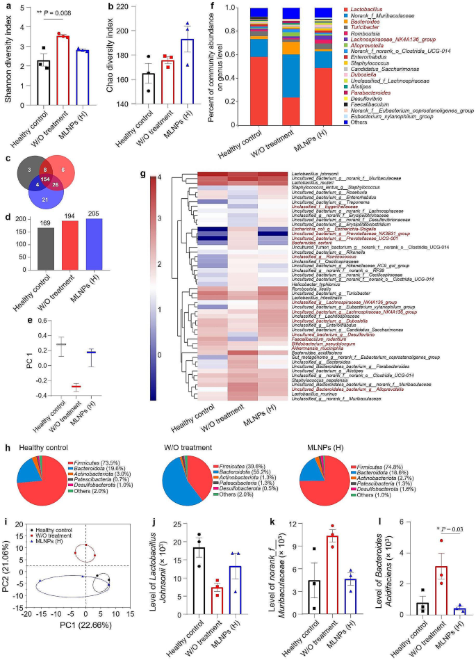
To additional elucidate the impacts of MLNPs on intestinal microbiota, we employed 16s RNA gene sequencing to comparatively examine the intestine microbiota compositions amongst totally different mouse teams. In contrast with the wholesome management group, DEN/NMOR remedy brought on a rise in bacterial richness on the species stage, accompanied by a corresponding elevation within the Shannon variety index representing microbiota variety. Oral MLNPs induced alterations within the microbiota diversities, as evidenced by adjustments within the Chao and Shannon variety index (Fig. 7a, b). Venn diagram can visualize the variety of frequent and distinctive species in addition to similarities and overlaps between totally different teams. It confirmed that the MLNP (H)-treated group had a outstanding improve in colony formation with a powerful depend of 205 strains (154 shared amongst all mouse teams and 21 distinctive to this group), which was in line with the development noticed within the Chao Index of Richness (Fig. 7c, d). Discrete statistics from principal coordinates evaluation (PCoA) illustrate how totally different teams are distributed alongside the PC1 axis. As introduced in Fig. 7e, the distances among the many wholesome management group, the DEN/NMOR management group, and the MLNP (H)-treated group mirrored the optimistic impact of MLNPs on microbiota reworking, demonstrating that MLNPs might successfully reverse intestinal flora issues attributable to chemical carcinogens whereas sustaining stability.
Analysis of reworking results of MLNPs on the intestinal microbiota. α-Diversities have been introduced by field plots of the (a) Shannon index, and (b) Chao index. Every level represents the imply ± S.E.M. (n = 3; *p < 0.05 and **p < 0.01). (c) Venn diagram of frequent and distinctive bacterial species. (d) Whole numbers of microbial species of mice in every group. (e) Dispersion of β-diversity from experiment end result. (f) P.c of neighborhood abundance on the genus stage of the intestinal microbiota. (g) Heatmap of relative abundance on the species stage of the intestinal microbiota. (h) Microbial compositions of varied mouse teams on the phylum stage. (i) Principal coordinates evaluation (PCoA) of the intestinal microbiota in every group. (j–l) Relative abundances of the standard useful and dangerous micro organism in numerous remedy teams. Every level represents the imply ± S.E.M. (n = 3; *p < 0.05 and **p < 0.01)
Moreover, the management group (with out remedy) exhibited a better relative abundance of detrimental micro organism (e.g., Norank Muribaculaceae and Bacteroides) when put next with the wholesome management group (Fig. 7f). Following oral remedy of MLNPs, there have been will increase within the relative abundances of Lactobacillus and Turicibacter. Earlier research have demonstrated that Lactobacillus can mitigate galactose-induced liver harm in rats by suppressing hepatic irritation, enhancing intestinal barrier perform, and modulating the regulatory metabolome of intestine microbiota [44]. Turicibacter belongs to the order Bifidobacteria, which regulates glycolipid metabolism as an intestinal probiotic. Moreover, it confirmed vital variations in a number of strains, together with Escherichia-Shigella, Uncultured bacterium Prevotellaceae NK3B31 group, and Uncultured bacterium Prevotellaceae UCG-001. These strains have been positively correlated with the management group (with out remedy) and negatively correlated with the wholesome management group and the MLNP (H)-treated group (Fig. 7g). As depicted in Fig. 7h, the Firmicutes stage decreased, whereas the degrees of Bacteroidota and Patescibacteria elevated within the DEN/NMOR management group. Within the intestine, Firmicutes and Bacteroidota are dominant strains for gram-positive and gram-negative micro organism, respectively. Adjustments of their ratios point out intestinal dysbiosis, which has been noticed in numerous liver ailments [45]. Research point out that almost all of pro-inflammatory micro organism are categorised as Patescibacteria, whereas quite a few probiotics fall below Firmicutes. Intestinal issues promote the proliferation of pro-inflammatory factor-producing micro organism and alter bile acid metabolism, thereby contributing to the event of HCC [46]. The MLNP (H)-treated group was capable of reverse the adjustments talked about above.
Research have demonstrated that Escherichia-Shigella, a member of the Enterobacteriaceae household, exerts its pathogenicity in nonalcoholic fatty liver illness sufferers by modulating lipid metabolism and selling lipid accumulation, finally leading to various levels of hepatic steatosis, irritation, and fibrosis [47]. The proportions of each Prevotellaceae and Bacteroides elevated in sufferers with superior liver cirrhosis [48]. Moreover, there was no overlap in colony construction between the wholesome mice and the DEN/NMOR management mice, with the latter being dispersed far aside. Nevertheless, after oral administration of MLNPs to liver most cancers mice, their colony constructions of the MLNP (H)-treated group partially intersected with these of the wholesome management mice (Fig. 7i). Consultant cultures have been chosen to showcase the trajectory of useful and detrimental micro organism following MLNP remedy (Fig. 7j-l). These outcomes collectively indicate that the remedy of MLNPs can effectively modulate the stability of intestinal flora.


