Supplies
3,4-dihydroxy-DL-phenylalanine (DL-DOPA), potassium permanganate (KMnO4), Mn ions requirements, 3-(4,5-dimethylthiazol-2-yl)-2,5-diphenyltetrazolium bromide (MTT), had been bought from Aladdin Reagent (Los Angeles, Southern California, USA). Thiol-terminated mannose-poly (ethylene glycol) (mannose-PEG-SH) was obtained from Xi’an ruixi Organic Technologu Co. Ltd. All of the chemical substances and supplies had been used as obtained with out additional purification except in any other case talked about. Deionized (DI) water (Millipore Milli-Q grade, 18.2 MΩ) was used all through the experiments. Clinically remoted extended-spectrum β-lactamase Escherichia coli (ESBL-producing E. coli), MDR Klebsiella pneumoniae (Ok. pneumoniae), MDR Pseudomonas aeruginosa (P. aeruginosa), and MDR Bacillus had been kindly supplied by Fuzhou Common Hospital. Methicillin-resistant Staphylococcus aureus (MRSA) was collected from The First Affiliated Hospital, Solar Yat-sen College.
Synthesis of MENP and MP-MENP
The MENP had been synthesized primarily based on our earlier report with minor modifications. Briefly, DL-DOPA (60 mL, 10 mM) suspension was launched right into a 100-mL flask and heated to 50 °C. Then, KMnO4 answer (1.8 mL, 100 mM) after ultrasonic dispersion was quickly added beneath vigorous stirring. This combination was maintained at 50 °C for six h to kind the MENP. To take away extra precursors and reactants, the obtained MENP had been washed 5 instances with deionized water through centrifugation (17,500 rpm, 15 min), and at last dispersed in water.
MP-MENP had been synthesized by mixing MENP with mannose-PEG-SH at a feeding mass ratio of 1:5 in alkaline buffer answer (pH ≈ 10.0). After vigorous stirring in a single day at room temperature, the merchandise had been washed a number of instances utilizing deionized water to take away residual mannose-PEG-SH. The ultimate MP-MENP had been dispersed in water and saved at 4 °C till use.
Characterization of MP-MENP
The hydrodynamic diameter and ζ potential had been detected by dynamic mild scanning (DLS) (NanoZS 90, Malvern, USA). Transmission electron microscopy (TEM, Tecnai G2 Spirit BioTwin, FEI, USA) was used to look at the morphology of MP-MENP. The focus of MP-MENP was measured utilizing UV − vis − NIR spectroscopy (Cary 5000, Agilent, USA) at 808 nm wavelength. The Mn content material was decided by inductively coupled plasma atomic emission spectroscopy (ICP-AES, Thermo) following digestion by aqua regia in a single day.
Photothermal impact of MP-MENP
On this research, 200 µL MP-MENP aqueous answer with totally different concentrations (0-200 µg/mL) was uncovered to a 808 nm laser (2.0 W/cm2, 5 min), and the pattern temperature was frequently recorded by an infrared thermal digital camera. Deionized water was set because the management.
Twin-modal imaging of MP-MENP
The PA alerts of MP-MENP had been collected utilizing a preclinical PA imaging system (Endra Nexus 128, Ann Arbor, MI) upon 808 nm laser excitation with a pulse width of seven ns and a repetition charge of 20 Hz. The reasonable laser vitality maintains at ∼5 mJ/cm2. The MRI relaxivity beneath varied magnetic fields was decided by a 7.0 T small animal MR scanner (Bio-Spec, Bruker, Karlsruhe, Germany), a 3.0 T medical scanner (Siemens, Prisma, Munich, Germany), and a 1.5 T HT-MICNMR-60 benchtop relaxometer (Huantong Company, Shanghai, China), respectively. The samples had been dissolved in water containing 1% agarose in Eppendorf tubes (1.5 mL).
In vitro anti-bacterial research on planktonic micro organism
5 clinically remoted bacterial strains, together with MRSA, MDR Bacillus, ESBL-producing E. coli, MDR Ok. pneumoniae, and MDR P. aeruginosa, had been employed in our experiment. The focus of micro organism was measured by the optical density at 600 nm through UV − vis − NIR spectroscopy. To evaluate the antibacterial capacity of MP-MENP, micro organism (106 CFU/mL) had been incubated with totally different concentrations of MP-MENP (6.25, 12.5, 25, 50 µg/mL). The micro organism with out MP-MENP co-incubation was used as management. The micro organism suspensions had been irradiated utilizing a 808 nm laser (2 W/cm2, 5 min). Then, the samples had been serially diluted. 100 µL of diluted bacterial suspensions was unfold on lysogeny broth (LB) agar plates and cultured for twenty-four h to look at the variety of bacterial colonies. The MP-MENP -treated micro organism with no laser irradiation had been counted following the aforementioned procedures. Every pattern was ready in triplicate.
Intracellular antibacterial exercise
RAW264.7 murine macrophage cells had been seeded on 96-well plates at a density of about 104 cells/effectively and contaminated with MRSA at a ratio of 10–20 micro organism per macrophage. To eradicate the extracellular micro organism, 50 µg/ml gentamycin was added into tradition media (DMEM). After in a single day, the MRSA-infected macrophages had been cultured in recent medium with MENP or MP-MENP at a focus of 100 µg/mL for 4 h. The group with out nanoparticles therapy was used as management. Then, the cell tradition medium was changed with recent medium, and the MRSA-infected macrophages had been irradiated at 808 nm, 2 W/cm2 laser for five min. The intracellular bacterial viability was decided by lysing contaminated macrophages in distilled water after which plating the lysates on LB plates for tradition and counting of bacterial colonies. The MRSA-infected cells with out laser irradiation had been counted following the aforementioned procedures. Every pattern was ready in triplicate.
For the dose-dependent antibacterial assay of MP-MENP, the MRSA-infected macrophage cells had been incubated with MP-MENP at totally different last concentrations (12.5, 50, 100, 200 µg/mL). After 4 h incubation, the combination was handled with or with out laser irradiation. Then, the cells had been lysed to isolate MRSA micro organism for colony counting. The cells with out MP-MENP therapy had been carried out as management.
In vitro PA and MR imaging
The MP-MENP at totally different last concentrations (50, 100, 200 µg/mL) had been incubated with RAW264.7 macrophage cells at 37 °C for 4 h. Subsequently, the cells had been collected and resuspended in 1% agarose for imaging analysis. The PA and MR photos had been recorded utilizing a 7.0 T small animal MRI scanner and NEXUS 128 scanner, respectively. The sign depth was decided by analyzing the area of curiosity.
Membrane integrity measurement
The MRSA had been cultured with 200 µg/mL MP-MENP. After laser irradiation, the micro organism had been remoted after which stained by SYTO 9 and propidium iodide (PI) for 30 min at nighttime. Afterward, micro organism had been washed with saline for a number of instances. Particular person suspensions had been immobilized on glass slides and imaged utilizing laser scanning confocal microscope (Olympus, FV1200).
Bacterial morphology research
The bacterial samples had been ready in keeping with the research of Dwell/Useless Bacterial Staining Assay. After pretreatment, the micro organism had been fastened utilizing paraformaldehyde for 4 h and noticed utilizing scanning electron microscope (SEM).
Western blotting
Equal volumes of MRSA from totally different teams had been collected and washed twice with PBS. After ultrasonication, bacterial sediments had been eliminated by centrifugation. The supernatant was incubated with 1% Protease Inhibitor Cocktail after which combined with a loading buffer adopted by heating at 100 °C for 10 min. Proteins from totally different teams had been added to SDS-PAGE and subsequently transferred to the poly(vinylidene difluoride) membrane. After being blocked, the membrane was consecutively incubated with antibodies in opposition to DnaK (1:1000 dilution) and GAPDH (1:10,000 dilution). The bands had been washed for HRP-conjugated secondary antibodies incubation. Lastly, the proteins had been imaged, and the chemiluminescence alerts had been detected.
Macrophage repolarization
RAW264.7 cells had been first pre-stimulated with 100 ng/mL of IL-4 for twenty-four h to polarize them into M2-type macrophages, after which co-incubated with LPS, MENP, or MP- MENP for an additional 24 h. The cell supernatant was rigorously collected for the cytokine secretion assay utilizing ELISA assay kits. In the meantime, the cells had been collected for APC-conjugated anti-mouse CD86 antibody (Biolegend, USA) and 0.5 µg PE-conjugated anti-mouse CD206 antibody (Biolegend, USA) incubation, after which detected utilizing stream cytometer.
Migration exercise of RAW264.7
The migration exercise of RAW264.7 was confirmed utilizing a transwell assay. Briefly, RAW264.7 cells (1 × 10⁵ cells/pattern) had been seeded onto 8 μm transwells. Subsequent, 2.5 mg/mL of MENP/MP-MENP had been added into the decrease chambers, whereas LPS and PBS had been additionally added into the opposite two teams as constructive and damaging controls. Following 24 h incubation, cells on the underside of the transwell had been stained with crystal violet and counted utilizing optical microscopy.
Phagocytic exercise of RAW264.7
RAW264.7 cells (2 × 10⁵ cells/effectively) had been seeded onto a 6-well plate and handled with PBS, LPS, MENP and MP-MENP teams, respectively. The assay for gentamicin safety was then carried out. After 24 h co-incubation, the supernatant was changed with recent DMEM containing MRSA (107 CFU/mL) for 30 min incubation. Then, the suspension was eliminated and new DMEM containing 200 µg/mL of gentamicin was added. Following 1 h tradition to eradicate the extracellular micro organism, cells had been washed with PBS totally. After that, 1 mL of 1% Triton X-100 was added to lyse macrophages, thereby releasing intracellular micro organism. Lastly, the Triton X answer from every effectively was collected to calculate the micro organism by gradient dilution and plate counting strategies.
Animal research
Male Balb/c mice (6 weeks, ~ 20 g) had been bought from Zhengzhou Hua-xing Laboratory Animal Heart. All animal research had been carried out in compliance with the protocols permitted by the Animal Administration and Ethics Committee of Henan College of Conventional Chinese language Medication. To evaluate the theranostic impact of MP-MENP, a mice mannequin of bacterial an infection was established by subcutaneous injection of MRSA micro organism (108 CFU/mL, 100 µL) into the correct thigh of mouse, whereas saline was injected to the left thigh as a management. After 24 h an infection the mice had been randomly divided into a number of teams (n = 5) for additional use.
In vivo imaging of MRSA an infection
The MRSA-infected mice had been injected with MENP or MP-MENP (at a dose of 20 mg/kg) through the tail vein, respectively. Within the case of aggressive inhibition experiment, the contaminated websites had been in situ injected with mannose to dam the infiltrated monocytes earlier than administrating MP-MENP. The PA photos from the MRSA-infected area at 0, 1, 3, 6, 12, and 24 h had been captured by FUJIFILM visualsonics (Toronto, Canada). The T1– and T2– weighted MR photos containing coronal planes had been acquired previous to and at 6 h postinjection of MP-MENP utilizing a 3.0 T medical scanner (Siemens, Prisma, Munich, Germany). The distinction enhancement was quantified by analyzing the ROI.
In vivo PTT of MRSA an infection
To guage antibacterial PTT efficacy of MP-MENP in vivo, mice with MRSA an infection had been randomly divided into 4 teams (n = 5): (1) saline with none therapy, (2) saline with 808 nm laser irradiation (2 W/cm2, 5 min), (3) MP-MENP alone, and (4) MP-MENP with 808 nm laser irradiation (2 W/cm2, 5 min). The laser publicity was carried out at 6 h post-injection. The contaminated areas had been recorded each 3 days. After 12 days of monitoring interval, the mice had been sacrificed, and the contaminated tissues had been harvested for hematoxylin and eosin (H&E) staining and CFU evaluation.
In vivo immunomodulation
After every therapy on day 2, mice had been sacrificed and the contaminated pores and skin tissues had been collected for immunofluorescence (IF) labeled with F4/80, CD86, CD206 associated antibodies. In the meantime, IF staining of HSP70, CD4+ T cells, and CD8+ T cells additionally carried out to confirm the immunological results. Furthermore, the collected pores and skin tissues had been additionally shortly frozen in liquid nitrogen on day 2, after which despatched to Novogene Co. Ltd. (China) for high-throughput sequencing evaluation. The enzyme-linked immunosorbent assay (ELISA) was launched to measure the serum cytokines in keeping with the protocols advisable by producer (ExCell Bio, Shanghai, China).
Security analysis
For cytotoxicity assay, human umbilical vein endothelial cells (HUVEC), RAW264.7 murine macrophage cells, and human hepatic cells LO2 had been used. The cells had been cultured in a single day on 96-well plates (10 000 cells/effectively), after which co-incubated with MP-MENP at totally different last concentrations (0, 50, 100, and 200 µg/mL) for twenty-four h. Subsequently, the cells had been washed 3 times utilizing PBS. The MTT answer (0.5 mg/mL, 10 µL) was added to every effectively. After 4 h incubation, the cell tradition medium was changed by 150 µL DMSO. The optical density of every effectively was decided at 490 nm utilizing a microplate reader.
Hemolysis evaluation of MP-MENP was examined on crimson blood cells (RBCs). The erythrocytes had been collected by centrifugation (1500 rpm, 15 min), after which washed 5 instances with saline. The centrifuged erythrocytes (3 mL) had been combined with saline (11 mL) to acquire the inventory dispersion of RBCs. Then, 100 µL inventory dispersion was added to saline answer (0.9 mL) containing MP-MENP at totally different concentrations. The ultimate RBCs stage was about 4%. Hereinto, deionized water and saline had been carried out because the constructive and damaging controls, respectively. After 3 h incubation at 37 ℃, the samples had been centrifuged at 12,000 rpm/min for 15 min. The share of hemolysis was decided by UV − vis evaluation of the supernatant at 540 nm absorbance, and calculated utilizing the next formulation:
% hemolysis = ((AS – AN) / (AP – AN)) ×100%
AS represents the absorbance of MP-MENP in RBCs suspension, AN represents the absorbance of RBCs suspension with saline therapy, and AP is the absorbance of pattern handled with deionized water.
For in vivo security analysis, wholesome Balb/c mice (6 weeks, ~ 20 g) had been randomly divided into two teams (n = 5) for saline or MP-MENP (20 mg/kg) administration. After 1-day postinjection, the mice had been sacrificed. The key organs together with coronary heart, liver, spleen, lung, and kidney had been collected and stuck utilizing 4% paraformaldehyde answer for H&E staining.
Statistical evaluation
Information evaluation was carried out with the GraphPad Prism software program. All information on this work had been introduced as imply ± SD. Statistical significance was decided by a One-way evaluation of variance (ANOVA). * means P < 0.05, ** means P < 0.01, and *** means P < 0.001.
Outcome and dialogue
Characterization of MP-MENP
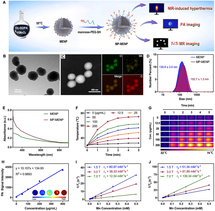
We first fabricated water-dispersible manganese-eumelanin coordination nanoparticles (MENP) by a one-pot intrapolymerization doping (IPD) technique (Fig. 2A). After easy chemical oxidation-polymerization of the DL-DOPA precursor with KMnO4, MENP had been efficiently obtained with a Mn loading effectivity of 8.2% wt/wt. Subsequently, mannose-terminal PEG was launched to switch the pristine MENPs for macrophage concentrating on. Noticed by transmission electron microscopy (TEM), the mannose-decorated MENPs (outlined as MP-MENP) had a well-controlled spherical morphology (Fig. 2B). The presence of oxygen, carbon, and manganese in MP-MENP was revealed by the excessiveangle annular darkish area scanning transmission electron microscopy vitality dispersive X-ray spectroscopy (HAADF-STEMEDX) mapping. The typical diameter of MP-MENP was about 152.7 nm as proven by dynamic mild scattering (Fig. 2D), which is barely bigger than pristine MENPs. The floor zeta potentials of MENPs and MP-MENP had been − 30.7 and − 35.8 mV, respectively. The variations of dimension and zeta potential between MENP and MP-MENP could also be attributed to the presence of hydration shell shaped between ample hydroxyl teams in mannose-terminal PEG and surrounding water molecules, which might probably stabilize the nanoparticles in addition to change their obvious properties.
(A) Schematic illustration of the preperation and performance of MP-MENP. (B) TEM picture and (C) HAADF-STEM-EDX mapping of MP-MENP. (D) Dimension distribution and (E) UV-vis-NIR − vis spectra of MENP or MP-MENP in deionized water. (F) The temperature variation and (G) thermographic photos of MP-MENP upon 808 nm laser irradiation (2 W/cm2, 5 min). (H) PA imaging alerts of MP-MENP with totally different concentrations. The inset exhibits the corresponding PA photos of MP-MENP. The linear relationship for the (I) r1 and (J) r2 relaxivities of MP-MENP as a perform of Mn focus
Photothermal and imaging results of MP-MENP
Photothermal efficiency is the important thing issue dominating the effectivity of antibacterial impact and subsequent immune activation. From the UV-vis-NIR absorption spectrum evaluation (Fig. 2E), there was no apparent NIR absorption change between MENP and MP-MENP within the NIR area, indicating that the potential photothermal property of nanomaterials won’t be impaired by mannose PEGylation. Determine 2F exhibits a gradual rise of temperature alerts with rising MP-MENP uncovered to laser irradiation (808 nm). Because the irradiation time extended, the temperature of MP-MENP underwent two phases (i.e., a gradual rise and leveling off) in succession (Fig. 2F, G). When arriving at steady-state, the temperature of MP-MENP aqueous answer might keep above 50 °C, regardless that its focus is low as 25 µg/mL. Such glorious photothermal impact is very conducive to additional organic software. We then investigated the aptitude of MP-MENP as a PAI and MRI distinction agent. As anticipated, the PA alerts of MP-MENP introduced a gradual enhancement because the focus elevated (Fig. 2H). Equally, a distinguished concentration-dependent distinction enhancement was noticed in each T1WI and T2WI., and the relaxivity variations at totally different magnetic fields (from 1.5 to 7.0 T) induced an apparent improve within the ratio of longitudinal (r1) to transverse (r2) relaxivity (Fig. 2I, J). All of those prompt that the MP-MENP might function a possible nanoplatform for imaging visualization and photothermal ablation of bacterial an infection.
In vitro antibacterial exercise on planktonic strains
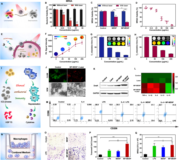
For antibiotic-resistant micro organism, regionally rising temperature (50 °C) by photothermal brokers will trigger an incredible inhibition on microbial metabolism, culminating in cell dying when in depth sufficient [20]. To guage the antibacterial photothermal exercise of MP-MENP, 5 clinically remoted MDR strains had been chosen (Fig. 3A). Amongst them, MRSA and MDR Bacillus are labeled as Gram-positive micro organism, and three different strains, together with ESBL E. coli, MDR Ok. pneumoniae and MDR P. aeruginosa, are the well-known Gram-negative species. The killing or antibacterial efficacy of MP-MENP at totally different concentrations was assessed by their corresponding bacterial viability utilizing colony counting methodology. As proven in Fig. 3B and S1, the viability values had been above 90% for all bacterial strains within the absence of NIR irradiation, indicating an nearly negligible antibacterial efficacy of the MP-MENP itself. Against this, the teams containing MP-MENP and NIR publicity confirmed apparent development inhibition, and the bacterial viability regularly decreased to lower than 5% with the rise of MP-MENP focus to 50 µg/mL. The affect of NIR mild could possibly be additionally excluded as no vital variations on the viability had been discovered at 0 µg/mL between the above two checks. Strikingly, the photothermal toxicity of MP-MENP was broadly efficient in each Gram-positive micro organism and Gram-positive species. It’s typically identified that treating infections brought on by MDR organisms, significantly their Gram-negative species, is a major problem for medical practitioners and vastly will increase affected person mortality and value of care globally. Contemplating the broad-spectrum photothermal harm, our developed MP-MENP might present a novel-acting strategy for sterilization no matter bacterial species and drug-resistance.
(A) Schematic illustration of MP-MENP mediated PTT in opposition to planktonic micro organism. The bacterial viability of (B) MRSA versus the MP-MENP concentrations with/with out laser irradiation (808 nm, 2 W/cm2, 5 min). (C) The micro organism viability of MRSA with or with out PTT by saline, MENP, and MP-MENP. (D) Focus-dependence of MRSA survival with MP-MENP upon laser irradiation (808 nm, 2 W/cm2, 5 min). (E) Schematic illustration of the theranostic MP-MENP for intracellular MRSA. (F) PA alerts, normalized (G) T1 and (H) T2 rest instances of MRSA-infected macrophages after co-incubation with MP-MENP. The insets in panels (F), (G), and (H) present corresponding PA, T1WI, and T2WI photos, respectively. (I) Schematic illustration of immuno-modulation impact by MP-MENP. (J) Consultant overlapping fluorescence photos for a stay/lifeless bacterial staining assay (sacle bar: 10 μm) and SEM photos (sacle bar: 500 nm) of MRSA with or with out MP-MENP-mediated PTT. (Ok) Western blot of HSP70 expression by RAW264.7 macrophages with totally different therapies. (L) ELISA outcomes of cytokines secreted by RAW264.7 after totally different therapies. (M) Detection of macrophage floor markers CD86 (M1 macrophage marker) and CD206 (M2 macrophage marker) utilizing a stream cytometer. (N) Schematic illustration of the transwell co-culture system. (O) Typical photos of RAW264.7 cells cultured on transwell stimulated with totally different conditional media. (P) Cell depend outcomes of the migrated RAW264.7 on the underside of the higher chamber of transwells in numerous circumstances. (Q) Counting outcomes of phagocytized MRSA by RAW264.7 handled in numerous teams. * means P < 0.05, ** means P < 0.01, and *** means P < 0.001
In vitro antibacterial exercise on intracellular MRSA
Intracellular infections are an impermeable barrier hindering the efficient administration of bacterial theranostics. When an infection does happen, monocytes not solely can chemotactically migrate to phagocytose invading organisms, but in addition function a possible shelter for micro organism, defending them from assaults by antibacterial medication. As soon as internalized by migratory monocytes, pathogens can survive inside cells for prolonged intervals and will attain distant websites to induce an infection, which might lead to long-term continual or recurrent instances [21, 22]. In our research, the MP-MENP had been functionalized with mannose which might goal monocytes after which induce receptor mediated endocytosis. In consequence, the micro organism hiding inside monocytes are expectantly eradicated by MP-MENP-mediated PTT. To confirm this speculation, RAW 264.7 was chosen because the mannequin monocyte host for MRSA invasion. The surviving intracellular micro organism after totally different therapies had been remoted and examined utilizing colony counting methodology. As famous, NIR irradiation alone didn’t trigger any viability inhibition on intracellular MRSA, whereas the micro organism handled with MENP confirmed exceptional development inhibition upon NIR activation (Fig. 3C). Making the most of macrophage-targeting capacity, the mannose-modified MP-MENP carried out probably the most environment friendly photothermal inactivation of intracellular MRSA, and such antibacterial efficacy was concentration-dependent (Fig. 3D). Nearly all of the intracellular MRSA had been killed on the MP-MENP focus of 200 µg/mL.
In vitro imaging of monocytes by MP-MENP
To verify that MP-MENP might efficiently lighten macrophages, the RAW 264.7 macrophage cells had been incubated with MP-MENP for 4 h, after which collected for PAI and MRI investigation (Fig. 3E). Mobile PA photos demonstrated incubation concentration-dependent sign enhancement (Fig. 3F). The harvested macrophages additionally confirmed apparent constructive and damaging distinction enhancement on T1WI and T2WI, respectively. Each the T1 and T2 rest time of cells had been regularly decreased because the incubation focus of MP-MENP improve (Fig. 3G, H). These outcomes elucidated that MP-MENP could possibly be successfully internalized by macrophages after which implement the dual-modal imaging of the cells. Additional coupled with potent photothermal ablation impact, MP-MENP expectantly act as a promising nanotheranostic to visualise an infection and implement a broad-spectrum killing of each planktonic micro organism and intracellular strains.
In vitro immunomodulation impact of MP-MENP
To confirm the proposed immunomodulation route of MP-MENP (Fig. 3I), SYTO 9 and PI co-staining was firstly launched, wherein the inexperienced fluorescent SYTO 9 was used to label stay micro organism, and crimson dye of PI permeates solely the broken bacterial cell membrane and has been proposed as a typical marker to analyze membrane integrity. As proven in Fig. 3J and S2, most of MRSA in management group introduced inexperienced shade through SYTO 9 staining, whereas the crimson PI fluorescence from broken micro organism was extensively present in MP-MENP-mediated PTT group. Additional noticed by SEM, the untreated MRSA micro organism had integral and easy our bodies (Fig. 3J). After publicity to MP-MENP and NIR laser, the bacterial morphology was dramatically modified, suggesting a sturdy bacterial disruption by MP-MENP-mediated PTT. The wrinkled, collapsed and even lysed cell partitions will result in the leakage of intracellular content material for micro organism. Together with lifeless micro organism and particles, such PAMPs are promising to activate potential immune response [23, 24]. Contemplating the hyperthermia-induced bactericidal impact of MP-MENP, we then investigated the expression of heat-shock proteins (HSPs) by MRSA after PTT, as a result of HSPs are consultant of defense-related proteins that resist thermal stress. As hazard alerts, they’re generally present in nearly all organisms, from micro organism to people, and may be quickly produced and launched when cells undergo from elevated environmental temperatures [25, 26]. From Fig. 3Ok, the DnaK (a classical HSP70 homologue of Staphylococcus aureus) was considerably upregulated in micro organism handled with MP-MENP plus laser irradiation, which implies an elevated expression of HSP70. As a well known ICD-related DAMPs, HSP70 has been extensively reported with a capability to set off immune response. Subsequently, the upregulation of HSP70 expression after MP-MENP-mediated PTT exhibits nice promise in enhancing antibacterial immunity.
The degrees of inflammatory cytokines following totally different therapies in RAW 264.7cells had been additional measured by ELISA equipment. As proven in Fig. 3L, the MP-MENP might considerably increase the secretion of M1-associated pro-inflammatory cytokines, together with tumor necrosis issue (TNF)-α, interleukin (IL)-1β, interleukin (IL)-6, and interleukin (IL)-12. In distinction, the M2-associated anti-inflammatory cytokine of interleukin (IL)-10 was clearly downregulated by MP-MENP stimulation. Within the case of bacterial an infection, the immunosuppressive microenvironment will trigger a change of host infection-associated macrophages, from pro-inflammatory macrophages (M1) to anti-inflammatory macrophages (M2) as a way to evade immunologic eradication. The M1-type macrophages couldn’t solely phagocyte and kill pathogenic micro organism straight, but in addition secret inflammatory elements to induce DCs maturation and thus reverse immune suppression [27, 28]. In consequence, repolarizing macrophages into M1 phenotype is taken into account as a promising therapeutic technique for bacterial an infection. To guage the repolarization functionality of MP-MENP on macrophages, IL4-was used to stimulate RAW264.7 macrophages (M2), and lipopolysaccharide (LPS) was launched as a constructive management to repolarize M2 macrophages into the basic inflammatory M1 inhabitants. The proportions of CD86 (M1 marker) and CD206 (M2 marker) macrophages had been assessed by stream cytometry. Determine 3M revealed that the proportion of M1-type macrophages elevated vastly (25.1%) within the IL-4 + MP-MENP group in contrast with the PBS alone (0.26%) and IL-4 group (0%). Furthermore, there may be few variations between the MENP and MP-MENP teams, which may be ascribed to the improved macrophage concentrating on and internalization supplied by mannose modification. Such M1-type polarization and pro-inflammatory cytokines secretion are conducive to reactivate antibacterial performance of macrophages, thereby reversing the immune microenvironment suppressed by pathogenic micro organism.
We then investigated the in vitro migration of macrophages utilizing the transwell assay (Fig. 3N). It was noticed that RAW264.7 cell migration on transwell was considerably enhanced within the presence of MP-MENP, which might permit macrophages to maneuver quickly to the an infection website (Fig. 3O, P). To additional consider the affect of nanoparticles on the anti-bacterial phagocytic capability of macrophages, RAW264.7 cells after totally different therapies had been co-cultured with MRSA. As proven in Fig. 3Q, the phagocytic variety of MRSA within the MENP group was way more than that within the PBS group, and the modification of mannose additional promoted the phagocytosis of macrophages. Collectively, MP-MENP was useful to reactivate antibacterial capabilities of macrophages, thus assuaging the immunosuppressive microenvironment of bacterial an infection.


