Synthesis and characterization of PEI-PTX (PP) polymers
The process for synthesizing PP polymers is proven in Further file 1: Determine S1. PTX-SA was obtained by conjugating succinic acid to the two’-OH group of PTX through ester bond formation. As confirmed in Further file 1: Determine S2, the huge and robust peak of 3438.38 cm−1 within the spectra of PTX-SA was originated from the hydrogen bond affiliation hydroxide, suggesting the existence of succinate. The 1H NMR spectrum and mass spectrum of PTX-SA was confirmed in Further file 1: Determine S3B and S4. HRESI-MS: m/z 976.72 [M + K]+, C51H55NO16. 1H NMR (400 MHz, DMSO-d6), δ (ppm): 12.28 (1H, s, b-COOH), 9.22 (1H, d, J = 8.5 Hz, 3’-NH-), 7.99–7.19 (15H, Ar–H), 6.29 (1H, s, 10-H), 5.81 (1H, t, J = 8.9 Hz, 13-H), 5.54 (1H, t, J = 8.7 Hz, 3’-H), 5.34 (1H, d, J = 9.0 Hz, 2-H), 4.93 (1H, m, 5-H), 4.64 (1H, s, 2’-H), 4.12 (1H, q, J = 6.8 Hz, 7-H), 4.02 (2H, q, J = 8.3 Hz, 20-H), 3.57 (1H, d, J = 7.1 Hz, 6-αH), 2.62 (2H, t, J = 6.4 Hz, a-H), 2.32 (2H, m, b-H), 2.24 (3H, s, 4-COCH3), 2.11 (3H, s, 10-COCH3), 1.87–1.79 (2H, m, 14-H), 1.76 (3H, s, 18-H), 1.63(1H, m, 6-βH), 1.50 (3H, s, 19-H), 1.02 (3H, s, 17-H), 1.00 (3H, s, 16-H). In the meantime, in contrast with the 1H NMR spectrum of PTX (Further file 1: Determine S3A), the height assigned to be the hydroxyl protons contributing to the two’-OH teams at δ 6.16 disappeared, which demonstrates the complete derivatization of all the two’-OH teams.
Then, PP was synthesized through amide response. The depth of infrared absorption peak, typically, is constructive associated with the change of dipole second. Within the spectrum of PEI-PTX (Further file 1: Determine S5), the height at 711.12 cm−1 was assigned to arene’s γCH of PTX, the height at 1245.07 cm−1 was assigned to oxhydryl’s υCO of PTX, and the height at 1112.84 cm−1 and 1069.34 cm−1 have been assigned to main (and secondary) amine’s υCN of PEI. The peaks at 1580.97 cm−1 and 1461.58 cm−1 within the spectrum of PEI have been assigned to main (and secondary) amine’s δNH of PEI. Nevertheless, the peaks at 1580.97 cm−1 and 1461.58 cm−1 within the spectrum of PEI weren’t proven within the spectrum of PEI-PTX. The first amine of PEI have been shift to amide, main the height at 1580.97 cm−1 being blue shifted and built-in to the peaks at 1648.84 cm−1. As well as, as a result of the secondary amine hydrogens of PEI have been fashioned hydrogen bonds in the course of the synthesis course of, the height at 1461.58 cm−1 was pink shifted and built-in to the peaks at 1245.07 cm−1. Within the spectra of PTX-SA, the absorption peak at 1722.69 cm−1 have been assigned to amide’s and ester’s υC=O, and the absorption peak at 1641.02 cm−1 was assigned to amide’s δNH. As well as, The N–H bending vibration (δNH) is a vital attribute of amine. After the amide response between carboxyl of PTX-SA and first amine of PEI, the depth of amide’s δNH peak at 1648.84 cm−1 was elevated due to the technology of latest amide. Thus, the FT-IR outcomes present the amide response between PTX-SA and PEI was efficiently synthesized. In addition to, in line with 1H NMR (400 MHz, DMSO-d6) (Further file 1: Determine S6), the peaks of δ 2.40 and δ 2.54 have been assigned to PEI, and the peaks of PTX in PP have been assigned as following: 9.22 (1H, d, J = 9.0 Hz, 3’-NH-), 8.05–7.10 (15H, Ar–H), 6.30 (1H, s, 10-H), 5.90 (1H, t, J = 5.9 Hz, 13-H), 5.56 (1H, m, 3’-H), 5.41 (1H, m, 2-H), 4.92 (1H, d, J = 4.9 Hz, 5-H), 4.62 (1H, m, 2’-H), 4.11 (1H, m, 6-αH), 4.01 (2H, dd, 20-H), 2.23 (s, 4-COCH3), 2.12 (s, 10-COCH3), 1.80 (1H, m, 14-H), 1.71 (3H, s, 18-H), 1.65(1H, m, 6-βH), 1.51 (3H, s, 19-H), 1.02 (6H, s, 16,17-H). The δ 9.05 within the 1H NMR spectrum of PP was originated from 3’-NH- in PTX, which was decrease than of PTX-SA (δ 9.22) as a result of PEI weakened the deshielading impact of succinate. In the meantime, there isn’t a peak assigned to 2’-OH at δ 6.16, which supported the conjugation between PTX and PEI through 2’-ester bond. The PTX content material of PP was decided in line with the 1H NMR spectra by evaluating integrals of peaks at δ 8.05–7.15 (fragrant protons of PTX) with δ 2.45–2.35 (partial methylene protons of PEI), which indicated that PP had 27.43 wt% PTX (calculation technique confirmed in Further file 1: Determine S6). GPC measurement confirmed that PTX was covalently bonded with PEI to kind polymer-drug conjugates (Further file 1: Determine S7). Nevertheless, there are the bimodal distribution and of PP indicated that a number of PTX can be grafted into PEI, and the cross-linking of PP might have triggered the distribution at low elution volumes.
Preparation and characterization of PP/siRNA/HA nanoassemblies
The method of self-assembly was proven in Fig. 2A. Initially, the PP copolymer dissolved in Milli-Q water could be self-assembled into micelles underneath sonication. Dynamic gentle scattering (DLS) evaluation confirmed that the hydrodynamic diameter of PP micelles was 215.7 ± 2.4 nm (Fig. 2F), and the transmission electron microscopy (TEM) photographs confirmed that the micelles have been spherical (Fig. 2E). As proven in Further file 1: Determine S7, the crucial micelle focus (CMC) of PP micelles was 6.3 × 10⁻³ mg/mL, indicating the superb thermodynamic stability of micelles. To find out the optimum weight ratio (PP/siRNA) for siRNA supply, we used agarose gel electrophoresis to judge the siRNA encapsulation of PP (Fig. 2B). It was discovered that binding capability elevated with growing the ratio of PP, and all of siRNA have been encapsulated when the burden ratio of PP: siRNA elevated to three:1. The encapsulation effectivity of siRNA was as much as 92.68 ± 0.78% at 3:1 weight ratio and maintained unchanged when additional growing ratios (Further file 1: Determine S9). As proven in Fig. 2C, an growing tendency of zeta potential was detected in the course of the enhance of weight ratios, additional indicating the profitable loading of siRNA. Due to this fact, the optimum weight ratio (PP/siRNA, w/w) of three:1 was used for additional anti-tumor research contemplating the encapsulation effectivity, z-average dimension, and zeta potential. The siRNA-loaded PP (PP/siRNA) confirmed a discount of constructive cost (from + 54.3 ± 1.6 mV of PP to + 47.5 ± 1.3 mV of PP/siRNA) and the slight lower of particle dimension (from 215.7 ± 2.4 nm.
to 197.3 ± 2.6 nm), indicating the condensing of siRNA on the PP floor (Fig. 2F). Subsequently, PP/siRNA was coated with a HA shell by electrostatic interplay. To find out the optimum ratio of HA and PP, we measured the z-average dimension and zeta potential of HA-coated PP/siRNA (PP/siRNA/HA) with completely different (HA: PP) ratios ranged from 1:2 to three:1. The floor cost of PP/siRNA/HA emerged reversal from constructive cost to detrimental when the burden ratio of HA: PP elevated to 2:1, and the particle dimension was decreased in the course of the enhance of HA ratio (Fig. 2D). Contemplating longer blood circulation and better tumor concentrating on, we used 2:1 as optimum weight ratio of HA/PP (w/w) for additional research. PP/siRNA/HA confirmed a gelatinous shell on floor in TEM (Fig. 2E), a reversal of zeta potential (from + 47.5 ± 1.3 mV of PP/siRNA to − 9.0 ± 0.3 mV of PP/siRNA/HA), and the lower of particle dimension (from 197.3 ± 2.6 nm of PP/siRNA to 144.4 ± 1.0 nm of PP/siRNA/HA) (Fig. 2F), revealing the presence of HA shell. The pictures of nanoassemblies PBS options have been confirmed in Further file 1: Determine S10, there have been translucent, azure, well-distributed colloidal options in PBS. To substantiate the drug loading contents (DLs) of PTX, completely different preparations have been hydrolyzed, after which the content material of PTX was measured by HPLC. The PP exhibited excessive drug loading effectivity (25.15 ± 0.90%, wt%), and the DLs of PP/siRNA, PP/HA, and PP/siRNA/HA have been 17.44 ± 0.43%, 7.27 ± 0.19% and 6.99 ± 0.15%, respectively (Desk 1). Notably, the PTX content material of PP decided by HPLC was barely decrease than by 1H NMR, which can be as a result of incomplete launch of PTX from PP micelles.
The PTX launch was investigated in vitro simulated surroundings. As proven in Determine S13 (A), the succinic acid ester in PP could possibly be hydrolyzed underneath acid situation or esterase catalysis. In contrast with pH7.4 group, PP might launch PTX on the presence of esterase, which indicated that PP would launch PTX by nonspecific esterase in cytoplasm. PP/siRNA/HA nanoassemblies remained secure and no drug leakage underneath pH5.0 and pH7.4, however hyaluronidase (1%, w/w) might promote the drug launch underneath acid situation, which indicated that hyaluronidase degraded HA shell. These outcomes demonstrated that the succinic ester in PP could be hydrolyzed by nonspecific esterase and launch of PTX in cytoplasm after PP escaping from endo/lysosomes to cytoplasm through the proton sponge mechanism.
Stability in vitro
The steadiness of nanoparticle system is important for its utility in vivo [42]. Each PP/siRNA and PP/siRNA/HA (containing 0.75 μM siRNA) in 50% FBS confirmed no free siRNA band upon gel electrophoresis (Fig. 2H). This outcome demonstrated that the HA shell contributed to the secure encapsulation of siRNA. To check whether or not the HA shell protects siRNA from anionic surroundings in physiological fluids and enzymatic problem in FBS, PP/siRNA and PP/siRNA/HA (containing 0.75 μM siRNA) have been subjected to 50% FBS. PP and PP/siRNA/HA remained secure for twenty-four h and confirmed no indicators of degradation as a result of presence of serum nucleases, whereas bare siRNA was utterly degraded in 12 h (Further file 1: Determine S11A). Heparin can displace siRNA from cationic gene carriers through ion alternate [43]. As proven in Determine S11B, PP/siRNA/HA confirmed stronger heparin resistance capability than samples with out HA-coated, additional supporting the protecting impact of the HA shell. Solubilizers, significantly cationic surfactants, often trigger extreme hemolytic response when injected into blood vessel [44]. Thus, we performed a hemolysis check to judge the protection of the PP and PP/HA. The PP trigger extreme hemolysis as we anticipated, however the PP/HA didn’t trigger hemolysis at a excessive focus and there was no important distinction when in comparison with the saline group (Fig. 2G and Further file 1: Determine S12), which demonstrated the HA shell not solely enhanced its stability, but in addition improved the protected and biocompatibility of nanoparticles. Thus, HA-coated PP could possibly be used for intravenous injection. Contemplating the extreme hemolysis and unsafety of PP with out HA-coated, we used the PP/HA or PP/siRNA/HA as most important preparations for additional anti-tumor research in vivo.
Cell uptake and endocytosis research
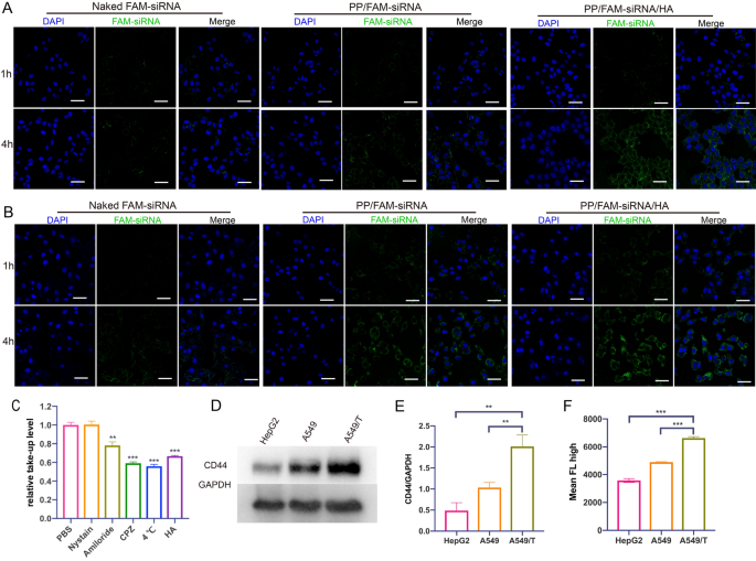
Environment friendly mobile uptake is important for gene transfection [41]. The confocal laser scanning microscope (CLSM) photographs of the FAM-siRNA (40 nM) in A549 and A549/T cells for 1 h and 4 h have been proven in Fig. 3A and B. Bare FAM-siRNA, in both A549 cells or A549/T cells, exhibited weak fluorescence depth at 1 h and 4 h since nucleic acid can hardly go via cell membrane [45]. The inexperienced fluorescence was clearly brighter after incubated with PP/FAM-siRNA or PP/FAM-siRNA/HA for 4 h, indicating siRNA-loaded nanoassemblies might promote gene internalization. Nevertheless, the fluorescence intensities at 4 h PP/FAM-siRNA/HA have been stronger than PP/FAM-siRNA as a result of PP/FAM-siRNA was unstable underneath wealthy serum surroundings. In the meantime, as incubation time was extended, the inexperienced fluorescence indicators grew to become stronger, indicating that extra nanoassemblies have been internalized into cells. This phenomenon was additionally verified by quantitative analysis by circulate cytometry (Further file 1: Determine S14). The outcomes confirmed that the PP/siRNA/HA might promote the uptake of siRNA in comparison with bare siRNA. Endo/lysosomal escape capability, avoiding the acid hydrolysis of gene, of FAM-siRNA loaded PP/siRNA/HA was evaluated by CLSM in both A549 or A549/T cells. As proven in Further file 1: Determine S15, we might observe that the FAM-siRNA loaded nanoparticles have been primarily entrapped in endo/lysosomal compartments at 1 h after uptake, whereas most FAM-siRNA might efficiently escape into the cytoplasm at 4 h. The colocalization analyzes and its Pearson’s coefficient have been additional verify this outcome. Due to this fact, the PP/siRNA/HA can exhibit glorious mobile internalization and endo/lysosomal escape capability, and could possibly be utilized in gene and protein drug supply.
Internalization of nanoassemblies. CLSM photographs of A549 cells (A) and A549/T cells (B) incubated with bare FAM-siRNA, PP/FAM-siRNA or PP/FAM-siRNA/HA nanoassemblies for 1 h and 4 h. Cell nuclei have been stained with DAPI. Scale bars: 50 μm (C) Endocytosis research of PP/siRNA/HA in A549/T cells by Stream cytometry (D) Western blot of P-gp expression in HepG2 cells, A549 cells, and A549/T cells. E Quantitive presentation of Western blotting. F Stream cytometry evaluation of uptake in HepG2 cells, A549 cells, and A549/T cells after handled with PP/FAM-siRNA/HA for 4 h. The info are offered as means ± SD. **p < 0.01 and ***p < 0.001
The internalization of PP/siRNA/HA nanocomplexes was investigated by A549/T cells following therapy with caveolin-mediated endocytosis inhibitors (nystatin), clathrin-mediated endocytosis inhibitors (chlorpromazine), macropinocytosis inhibitors (amiloride), and power inhibition (4 °C), respectively [46]. As displayed in Fig. 3C, the endocytosis of nanocomplexes was considerably inhibited underneath microthermal situation (4 °C), demonstrating the energy-dependent endocytosis mechanisms. The ends in Fig. 3C revealed that nanoparticle uptake was considerably inhibited by chlorpromazine and amiloride therapy, suggesting that clathrin-mediated endocytosis and macropinocytosis might each play a task within the uptake of PP/siRNA/HA. In the meantime, there may be an extra group of overdose HA blocking CD44-receptor to research the uptake of PP/siRNA/HA, indicating that the endocytosis can be CD44-mediated endocytosis pathway. Hyaluronic acid, as reported, can improve the internalization of nanocomplexes via binding CD44 receptor [47]. The CD44 expression of A549 cells, A549/T cells, and HepG2 cells (as CD44-negative cells) have been detected by Western blotting (Fig. 3D and E). This outcome demonstrates that the CD44 expression of A549/T cells was considerably increased than A549 cells and HepG2 cells. To judge the CD44-mediated internalization, we investigated the uptake of PP/siRNA/HA by circulate cytometry (Fig. 3F). The fluorescence depth in A549/T cells was statistically increased than A549 and CD44-negative HepG2, suggesting the excessive potential concentrating on capability of HA-coated nanocomplexes.
Silences gene expression in vitro
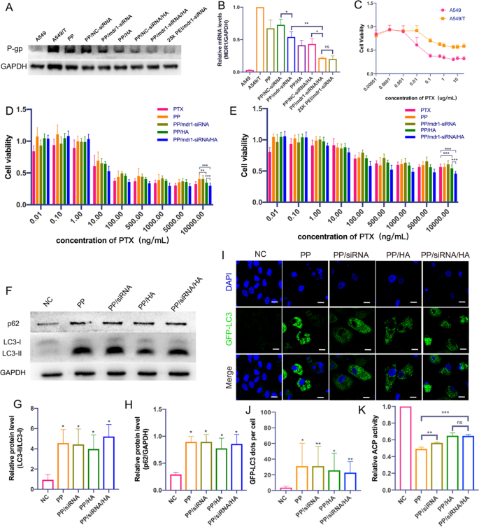
Evaluating to that on A549 cells, the P-gp on the membrane of A549/T cells was important over-expressed (Fig. 4A). 25 Ok b-PEI, as reported, was all the time used as constructive management on account of its glorious transfection effectivity [48]. After handled with PP/siRNA and PP/siRNA/HA, the quantity of P-gp on A549/T cells was considerably lowered. The silence effectivity was confirmed by quantitative PCR (Q-PCR) evaluation of transfected A549/T cells, the expressions of mdr1 have been lowered to 54% and 21% (Fig. 4A and B). There isn’t a important distinction in gene silencing between PP/siRNA/HA and 25 Ok PEI. Nevertheless, it was discovered that each PP and PP/HA, with out siRNA-loading, confirmed the suppression to mdr1 gene and decreased mdr1 expression to 67% and 41%, respectively. As well as, NC-siRNA loaded nanocomplexes (PP/NC-siRNA and PP/NC-siRNA/HA) didn’t lower the expression of P-gp in contrast with PP and PP/HA, which indicated the silencing impact of mdr1 siRNA was particular. Some earlier research confirmed that autophagy had an necessary position within the improvement of multi-drug resistance [49]. Moreover, some compounds like chloroquine or cationic polymers might block regular autophagy flux by alkalizing lysosomes [50]. Thus, we initially guessed that the gene suppression could also be brought on by autophagy modulation. As well as, it could possibly be defined for the instability of PP/siRNA with out HA-coated that the silence effectivity was decrease than PP/HA and PP/siRNA/HA.
Reversing MDR of nanoassemblies (A) Western blot of P-gp expression in A549/T cells after handled with PP, PP/NC-siRNA, PP/mdr1-siRNA, PP/HA, PP/NC-siRNA/HA, PP/mdr1-siRNA/HA and 25 Ok PEI/siRNA (B) Relative quantification of mdr1-gene expression by q-PCR evaluation. C Cytotoxicities of PTX in A549 and A549/T cells. Cell viability handled with numerous concentrations of PTX and nanoassemblies. D A549 cells, E A549/T cells. (F) Western blot of LC3 and p62 expression in A549/T cells after handled with nanoassemblies for 48 h. G, H Quantitative presentation of Western blotting. T check in comparison with NC group. I CLSM photographs of GFP-LC3 constructive dots in A549T/GFP-LC3 handled with nanoassemblies for 48 h. Scale bars: 20 μm. J Quantified results of GFP-LC3 constructive puncta. Ok The acid phosphatase exercise in A549/T cells by nanoassemblies for 48 h. The info are offered as means ± SD. *p < 0.05, **p < 0.01, and ***p < 0.001
Cytotoxicity and MDR-reversing of nanoparticles
The cytotoxicity ranges of various PTX formulations on A549 and A549/T cells have been decided by MTT assay [51]. A549/T cells have been chosen to check MDR-reversing as a result of it might overexpress P-gp, as talked about above, and exhibited resistance to PTX (Fig. 4C). LMW PEI had the benefit of low-cytotoxicity [52], 1.8 Ok PEI confirmed low cytotoxicity ranges in each A549 cells and A549/T cells (Further file 1: Determine S16). As proven in Fig. 4D and E, all completely different PTX formulations might cut back the viability of A549 and A549/T cells and exhibit concentration-dependent cytotoxic results. The outcomes confirmed each PTX formulations unfolded glorious anticancer results on A549 cells. The inhibition ratios in opposition to A549/T cells have been evidently decrease than that on A549 due to the drug-resistance of A549/T cells. Extra necessary, PP/siRNA/HA might extra successfully improve the inhibition ratio of A549/T cells than different PTX formulations. Notably, the superb anticancer impact of PP/siRNA/HA might counsel gene silencing the cytotoxicity ranges of various formulations on A549/T cells having relationship with the suppression of P-gp. Though PP might lower the expression of P-gp, the inhibition ratio of it was decrease than free PTX, which can be interpreted because the nanostructure disturbed by wealthy serum surroundings [41].
The half maximal inhibitory focus (IC50), decrease IC50 representing increased cytotoxicity, was additional used to confirm the cytotoxicity of the completely different formulations in opposition to most cancers cells. In the meantime, the IC50 worth is among the most necessary indexes about drug resistance of most cancers cells [9]. The IC50 values of various PTX formulations in opposition to A549 and A549/T cells have been listed in Desk 2. The IC50 of free PTX on A549/T cells (5.5 ± 0.5 ng/mL) was 16.5-fold over that on A549 cells (90.6 ± 14.0 ng/mL), indicating the A549/T cells had acquired an incredible drug-resistance. The IC50 values to A549 cells have been related and barely increased than that of free PTX, respectively. This was as a result of energetic ingredient (PTX) wanted launched from preparations [42]. The IC50 of PP, PP/siRNA, PP/HA, and PP/siRNA/HA in opposition to A549/T cells have been 35.5 ± 14.6 ng/mL, 77.5 ± 10.4 ng/mL, 31.5 ± 7.4 ng/mL, and 15.4 ± 3.0 ng/mL, respectively. PP/siRNA/HA exhibited higher cytotoxicity and reversal of drug resistance on A549/T cells than different formulations.
Cell apoptosis and cell cycle
To additional verify the therapeutic impact of various PTX formulations (containing 10 ng/mL of PTX) on A549 and A549/T cells, the cell apoptosis by Annexin V-FITC/PI staining was carried out on the similar concentrations of 10 ng/mL of PTX. As displayed in Further file 1: Determine S17A, each PTX formulations considerably induced late apoptosis in opposition to A549 cells. Nevertheless, for A549/T cells, free PTX couldn’t induce important apoptosis (Further file 1: Determine S17B). PP/siRNA/HA induced the very best apoptosis fee (87.9%) in opposition to A549/T cells in contrast with the management (6.7%), and PP/HA might induce related apoptosis fee (85.4%). Notably, PTX formulations primarily induced early apoptosis in opposition to A549 cells however, to A549/T cells, primarily late apoptosis. This may occasionally outcome from the upper tolerance of A549/T cells. As proven in Further file 1: Determine S18A and B, PTX might are inclined to arrest A549 cells in G2/M section. Nevertheless, the cell cycle of A549/T cells was not considerably completely different between PTX and controls, additional indicating the drug resistance of A549/T cells. PP/siRNA/HA might cut back G0/G1 section (from 65.7 to 34.7%) and have a tendency to arrest in S section (Further file 1: Determine S18C), suggesting the operate mechanisms of PP/siRNA/HA nanocomplex engaged on A549/T cells could be completely different from that of PTX due to autophagy inhibition. These outcomes confirmed the flexibility of PP/siRNA/HA to reverse drug resistance.
Autophagy modulation research
The gene silences experiments confirmed that each PP and PP/HA, with out siRNA-loading, might suppress mdr1 gene expression (Fig. 4A and B). To raised perceive the rationale that PP and PP/HA (containing 2 μM of PP) decreased mdr1 expression, the LC3/P62 expressions and numbers of autophag/autolysosomes have been used to evaluating their autophagy-modulated results. The image of autophagy flux initiation is the conversion of LC3B-I into LC3B-II, LC3B-I is free in cytoplasm however LC3B-II is instantly binding to autophagosomes membranes [53]. In the meantime, the extent of p62, as a degradation substrate of autophagy, can mirror the autophagy flux [54]. As illustrated in Fig. 4F–H, after therapy by similar gene silences experiments, there have been important accumulate of LC3-II and p62 in comparison with management group. Subsequently, the GFP-LC3 puncta have been noticed to dramatic will increase underneath CLSM (Fig. 4I and J). Our earlier analysis reveals that the talents of lysosomal acidity and blocking autophagic flux would mirror within the decreased exercise of acid phosphatase (ACP) [55]. As proven in Fig. 4Ok, each nanocomplexes might lower the ACP exercise. These outcomes prompt that nanocomplexes (PP, PP/siRNA, PP/HA, and PP/siRNA/HA) might induce autophagosome accumulation and block autophagic flux. Moreover, we preliminarily thought of that PP was the primary element to modulating autophagic flux.
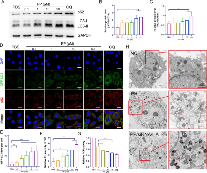
To additional confirm the autophagy modulation of PP, the markers of autophagy have been assessed by western blotting and CMSL. CQ, as a typical autophagy inhibitor, can block the fusion of autophagosomes through alkalizing lysosomes [56]. In our research, CQ (10 µM) can be acted as constructive management of autophagy inhibition. After handled with PP for twenty-four h, LC3-II/LC3-I ratio confirmed elevated and triggered the buildup of p62 (Fig. 5A–C). As confirmed in Fig. 5D and E, the GFP-LC3 dots per cell have been elevated from 7.4 to twenty-eight.5 because the focus enhance from 0.1 to 50 µM PP. The relative fluorescence intensities of p62 additionally have been elevated from 1.1 to 2.9 (Fig. 5F). The outcomes of relative ACP exercise confirmed that PP might alkaline lysosome and block degradation of autophagic substrates (Fig. 5G). TEM is a vital technique to look at the ultrastructural options of autophagosomes [57]. Bio-TEM photographs confirmed that upon comparisons with the management, many small double/multi-membrane vesicles and big vacuoles have been noticed after therapy of 100 µM PP or PP/siRNA/HA for twenty-four h (Fig. 5H). These outcomes demonstrated that prime dose of PP might block autophagic flux through alkalizing lysosomes.
Autophagy modulation of nanoassemblies (A) Western blot of LC3 and p62 expression after incubated with numerous concentrations of PP (0–50 μM), 10 μM CQ as constructive management. B, C Quantitative presentation of Western blotting. D CLSM photographs of GFP-LC3 constructive dots (inexperienced) and p62 immunofluorescence staining (pink) in A549T/GFP-LC3 incubated with numerous concentrations of PP. Cell nucleus have been stained by DAPI. Scale bars: 20 μm (E) Quantified results of GFP-LC3 constructive puncta. F The quantitative fluorescence depth of p62. G The acid phosphatase exercise after therapy of PP. H Bio-TEM photographs of A549/T cells after incubated with PP (50 μM) and PP/siRNA/HA (50 μM) for twenty-four h. The arrows point out the autophagosomes. The info are offered as means ± SD. *p < 0.05, **p < 0.01, and ***p < 0.001
In Vivo biodistribution
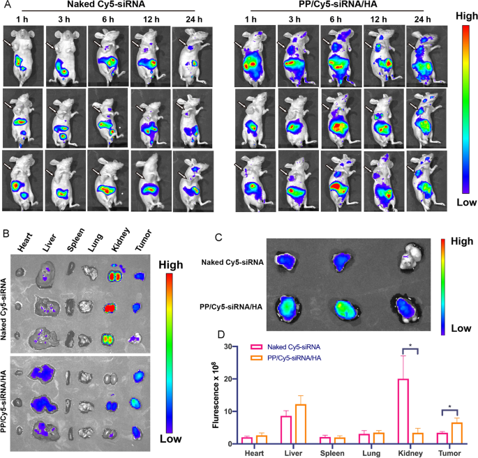
The bio-distribution of Cy5-siRNA loaded nanoassemblies was assessed by in vivo fluorescence imaging system after the tail vein injection of PP/Cy5-siRNA/HA in mice [58]. As proven in Fig. 6A–C, the fluorescence of bare Cy5-siRNA intensely distributed within the kidneys, however the PP/Cy5-siRNA/HA teams confirmed fluorescence primarily distributed within the liver, tumor, and kidneys. This could possibly be defined by that Cy5-siRNA in resolution could possibly be excreted by the kidneys however the nanoassemblies (~ 200 nm) could possibly be enriched in liver [59]. As well as, the fluorescence indicators arising from the tumors of mice handled with PP/Cy5-siRNA/HA was 1.9-fold increased than that handled with bare Cy5-siRNA (Fig. 6D). This means that HA-coated nanoassemblies can’t solely lengthen blood circulation time but in addition enrich on the stage of tumor tissue.
In vivo biodistribution of the PP/Cy5-siRNA/HA (1 mg/kg of Cy5-siRNA). A Photographs of whole-body after intravenous administration of Bare Cy5-siRNA or PP/Cy5-siRNA/HA in mice. The arrows point out the tumor location. B Photographs of extracted most important organs. C Photographs of extracted tumors alone. D Fluorescence effectivity of extracted most important organs at 24 h. The info are offered as means ± SD. *p < 0.05
In Vivo anti-tumor impact and RNAi effectivity
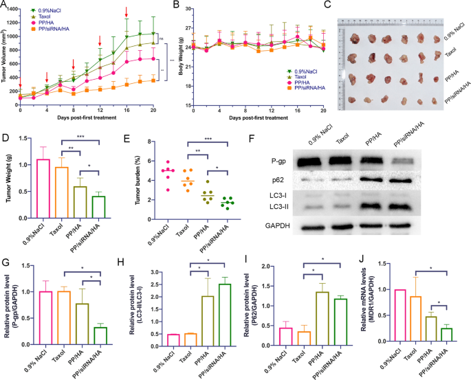
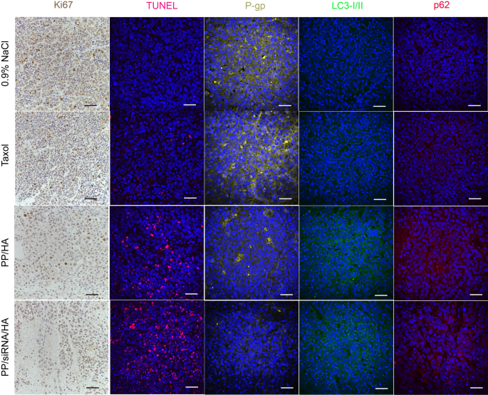
The antitumor impact was additional evaluated in A549/T tumor-bearing nude mice. As illustrated in Fig. 7A, there was no important distinction between 0.9% NaCl group and Taxol group. As compared, PP/siRNA/HA exhibited a superior antitumor effectivity with the tumor development inhibition charges of 36.43% (Fig. 7C–E). In contrast with the 0.9% NaCl and paclitaxel teams, the PP/HA group had enhanced therapeutic results, however was nonetheless inadequate to suppress drug-resistant tumors. The tumor tissue handled with PP/siRNA/HA exhibited a extra clearly decline in P-gp protein expression (Fig. 7G) and mdr1 gene expression (Fig. 7J) in contrast with different teams. The TUNEL outcomes confirmed that the PP/siRNA/HA successfully induced extra apoptosis within the tumor in contrast with Taxol and PP/HA (Fig. 8). The mobile proliferation of tumor was assessed by Ki67 assay (Fig. 8). In contrast with the opposite teams, the PP/siRNA/HA group had the least mobile proliferation and was discovered to have the most effective antitumor efficacy. The results of immunofluorescence staining additional confirmed the gene silence effectivity of PP/siRNA/HA with vastly lowered P-gp expression (Fig. 8). This demonstrated the PP/siRNA/HA nanocomplexes might efficiently reverse the drug-resistance of tumors, and realized tumor inhibition by gene/ drug co-delivery. The weights of all teams maintain principally untouched all through the therapy (Fig. 7B). There was no important physiological morphology abnormality in coronary heart, liver, spleen, lung, and kidney, confirming the protection for these samples (Further file 1: Determine S19).
In vivo therapy efficacy of nanoassemblies in opposition to A549/T xenograft tumors (n = 6). A The tumor development curves after handled with completely different formulations. The pink arrows point out the dates of administration. B Physique weight modifications. C Photos of extracted tumors after final therapy. D The weights of extracted tumors. E Tumor burden. F Western blot of P-gp, p62, and LC3 expression of the tumors. G–I Quantitative shows of Western blotting. J The mdr1-gene expression of the tumors by q-PCR evaluation. The info are offered as means ± SD. *p < 0.05, **p < 0.01, and ***p < 0.001
In vivo autophagy modulation
To additional verify the autophagy blockade impact of nanocomplexes, the expression of autophagy-associated proteins (LC3 and p62) in tumor was monitored on the finish of the remedies. The LC3B-I/II and p62 proteins expression in tumor tissue have been evaluated by western bolting and immunofluorescence staining. As confirmed in Fig. 7F–I, the LC3B-I/II and p62 proteins expression of PP/HA and PP/siRNA/HA have been increased than 0.9% NaCl and Taxol. Furthermore, there aren’t any important completely different between PP/HA and PP/siRNA/HA within the protein expressions of LC3B-I/II and p62. Nevertheless, in vivo, tumor microenvironment performs an necessary position within the improvement of tumor development and drug-resistance [51]. Thus, in contrast with PP/siRNA/HA, PP/HA simply performed restricted impact of anti-tumor in vivo. The outcomes of immunofluorescence staining additional confirmed the effectivity of blocking autophagic flux (Fig. 8).


