

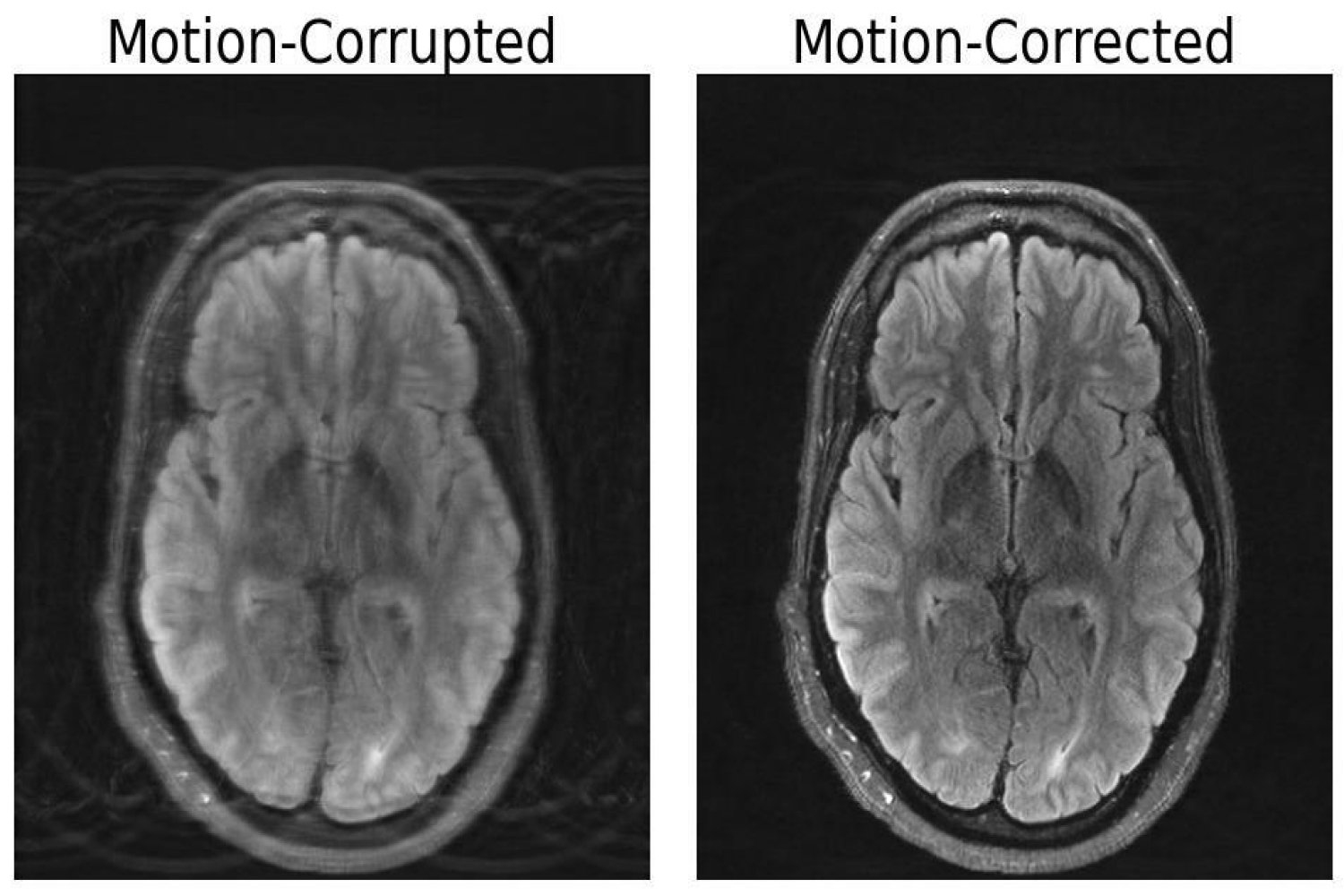
In comparison with different imaging modalities like X-rays or CT scans, MRI scans present high-quality delicate tissue distinction. Sadly, MRI is very delicate to movement, with even the smallest of actions leading to picture artifacts. These artifacts put sufferers liable to misdiagnoses or inappropriate therapy when vital particulars are obscured from the doctor. However researchers at MIT could have developed a deep studying mannequin able to movement correction in mind MRI.
“Movement is a standard downside in MRI,” explains Nalini Singh, an Abdul Latif Jameel Clinic for Machine Studying in Well being (Jameel Clinic)-affiliated PhD pupil within the Harvard-MIT Program in Well being Sciences and Expertise (HST) and lead writer of the paper. “It’s a reasonably sluggish imaging modality.”
MRI classes can take anyplace from a couple of minutes to an hour, relying on the kind of photographs required. Even throughout the shortest scans, small actions can have dramatic results on the ensuing picture. Not like digital camera imaging, the place movement sometimes manifests as a localized blur, movement in MRI typically ends in artifacts that may corrupt the entire picture. Sufferers could also be anesthetized or requested to restrict deep respiratory with a view to reduce movement. Nonetheless, these measures typically can’t be taken in populations significantly vulnerable to movement, together with youngsters and sufferers with psychiatric issues.
The paper, titled “Knowledge Constant Deep Inflexible MRI Movement Correction,” was not too long ago awarded greatest oral presentation on the Medical Imaging with Deep Studying convention (MIDL) in Nashville, Tennessee. The tactic computationally constructs a motion-free picture from motion-corrupted knowledge with out altering something concerning the scanning process. “Our goal was to mix physics-based modeling and deep studying to get one of the best of each worlds,” Singh says.
The significance of this mixed method lies inside making certain consistency between the picture output and the precise measurements of what’s being depicted, in any other case the mannequin creates “hallucinations” — photographs that seem reasonable, however are bodily and spatially inaccurate, probably worsening outcomes in terms of diagnoses.
Procuring an MRI freed from movement artifacts, significantly from sufferers with neurological issues that trigger involuntary motion, corresponding to Alzheimer’s or Parkinson’s illness, would profit extra than simply affected person outcomes. A examine from the College of Washington Division of Radiology estimated that movement impacts 15 p.c of mind MRIs. Movement in all forms of MRI that results in repeated scans or imaging classes to acquire photographs with ample high quality for analysis ends in roughly $115,000 in hospital expenditures per scanner on an annual foundation.
In response to Singh, future work may discover extra refined forms of head movement in addition to movement in different physique elements. For example, fetal MRI suffers from speedy, unpredictable movement that can not be modeled solely by easy translations and rotations.
“This line of labor from Singh and firm is the subsequent step in MRI movement correction. Not solely is it glorious analysis work, however I imagine these strategies will probably be utilized in every kind of scientific circumstances: youngsters and older of us who cannot sit nonetheless within the scanner, pathologies which induce movement, research of transferring tissue, even wholesome sufferers will transfer within the magnet,” says Daniel Moyer, an assistant professor at Vanderbilt College. “Sooner or later, I believe that it possible will probably be commonplace follow to course of photographs with one thing straight descended from this analysis.”
Co-authors of this paper embody Nalini Singh, Neel Dey, Malte Hoffmann, Bruce Fischl, Elfar Adalsteinsson, Robert Frost, Adrian Dalca and Polina Golland. This analysis was supported partly by GE Healthcare and by computational {hardware} supplied by the Massachusetts Life Sciences Middle. The analysis workforce thanks Steve Cauley for useful discussions. Extra help was supplied by NIH NIBIB, NIA, NIMH, NINDS, the Blueprint for Neuroscience Analysis, a part of the multi-institutional Human Connectome Venture, the BRAIN Initiative Cell Census Community, and a Google PhD Fellowship.
