CR-DPA-T synthesis and characterization
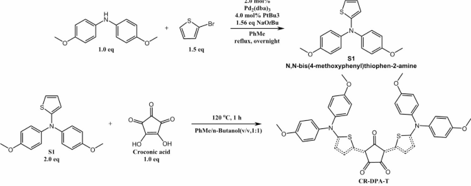
D-A-D kind CR-DPA-T was synthesized by a facile two-step response in keeping with beforehand reported methodology with commercially accessible supplies, as proven in Scheme 2 [35]. Condensation of electron-withdrawing croconic acid (A) with two equivalents of two sturdy electron-donating diphenylamine (DPA) models linked to thiophene linkers at 1:1 azeotropic combination of benzene and n-butanol at 110 o C, yielding CR-DPA-T as a black stable (28%). All spectral information of the intermediates (S1) and CR-DPA-T matched nicely with that of earlier report [34]. In the meantime, the D–π–A–π–D CR-DPA-T framework led towards appreciable electron delocalization and red-shifted absorption, leading to wonderful excessive chemical/thermal/picture stability [36, 37].
Characterization of EVs engineered with CDH17 nanobody
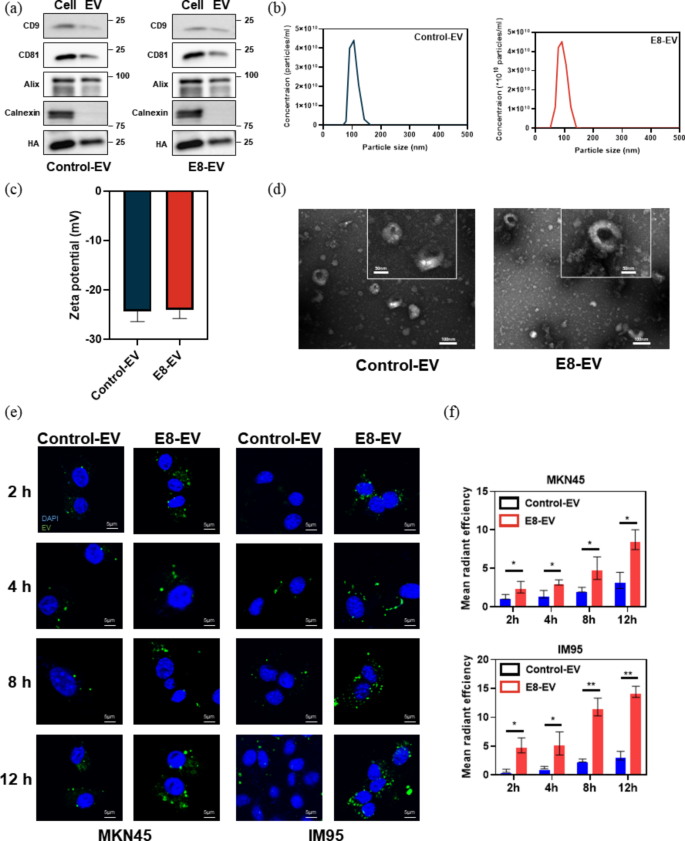
It was reported that CDH17 is a perfect diagnostic marker for GI tract cancers [29,30,31,32,33,34]. Our earlier work had confirmed the concentrating on and cell uptake means of EVs engineered with CDH17 nanobody E8 [34]. Right here, we recapitulated the traits of E8-EVs and management nanobody-EVs (Con-EVs) utilizing particular EVs optimistic biomarkers (CD9, CD81, Alix), along with unfavourable biomarker (Calnexin) (Fig. 1a). HA tag was detectable on the EVs, indicating the nanobodies fused with HA tag had been engineered onto EVs. Then, the EVs nanoparticle parameters had been decided by NTA. The typical measurement of the Con-EVs and E8-EVs was 103 ± 28 and 120 ± 16 nm, respectively (Fig. 1b), whereas the corresponding Zeta potentials (ζ) had been − 24.3 ± 3.8 and − 24.1 ± 3.3 mV, respectively (Fig. 1c). Each of the engineered-EVs are bodily comparable. TEM indicated that the diameters for essentially the most of particle EVs ranged from 30 to 120 nm, having negligible variations inside morphology (Fig. 1d).
To substantiate the specificity of E8-EVs in opposition to CDH17, we first knocked down the CDH17 protein in MKN45 cells with shRNA after which assessed the binding specificity of E8-EVs to MKN45 with/with out CDH17 knockdown. As proven in Fig S2, knockdown CDH17 virtually abolished the binding exercise of E8-EVs to CDH17-positive cells; nonetheless, management shRNA which can’t disturb the expression of CDH17 didn’t affect the binding functionality of E8-EVs to MKN45 cells. These information point out that E8-EVs can particularly work together with CDH17 protein expressing on the floor of most cancers cells. Subsequent, to additional show that EVs could possibly be effectively internalized by CDH17 optimistic tumor cells, PKH67-labeled EVs had been incubated with two gastric most cancers cell traces MKN45 and IM95 at numerous time factors, each of which had been confirmed excessive expression of CDH17 protein [33]. Immunofluorescence staining revealed that EVs could possibly be successfully internalized in MKN45 and IM95 cells in a time-dependent method; nonetheless, fluorescence depth of E8-EVs was superior to manage EVs, which implied that internalization was enhanced because of the interplay between E8 nanobodies and membrane CDH17 protein. (Determine 1e and f). These outcomes reveal that E8-EVs are able to bettering the concentrating on capability in the direction of CDH17-overexpressing cells, due to this fact selling mobile uptake.
(a) Western blot evaluation of particular EV biomarkers and HA tag. (b) NTA for EVs. (c) The zeta potentials for EVs. (d) TEM picture for EVs. Inset is the magnified picture of EVs. Scale bars, 100 nm and 50 nm respectively. (e) The internalization effectivity of EVs labeled with PKH67 (inexperienced) within the CDH17-positive MKN45 and IM95 cells beneath completely different time factors. Scale bars, 5 μm. (f) The quantification of internalization of EVs for e. Information are expressed as imply ± SEM
Characterization of photophysical properties of CR@E8-EVs
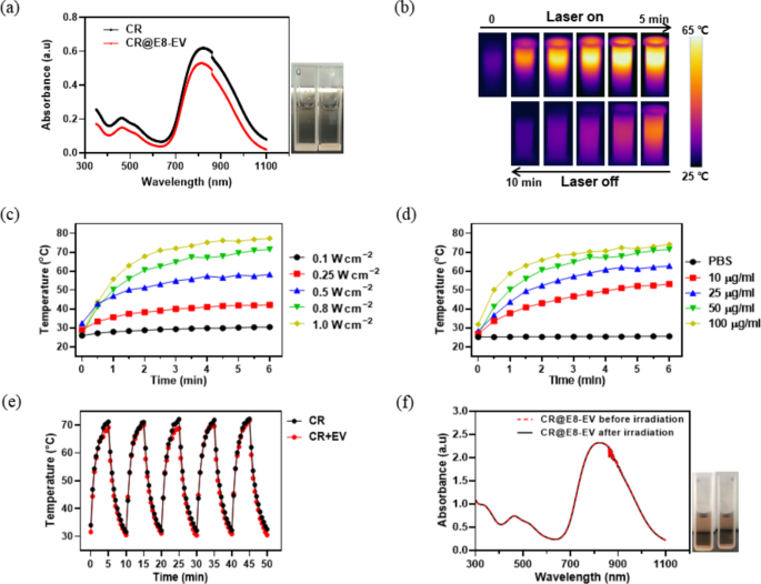
Subsequent, we encapsulated CR-DPA-T into the E8-EVs as described within the half 2.3, which was termed as CR@E8-EVs. To show that EVs don’t change the intrinsic traits of CR dye, we investigated the photophysical properties of CR@E8-EVs in answer state. As examined inside UV–vis–NIR absorption spectrum (Fig. 2a), free CR and CR@E8-EVs displayed comparable absorption sample (400 ~ 1100 nm) centered at roughly 810 nm, matching nicely with the 808 nm laser. There had no distinction of the absorption and shade between the free CR and CR@E8-EVs (inset photographs in Fig. 2a). The quantity and loading effectivity of CR encapsulated within the ready CR@E8-EVs was decided by the calibration absorption curve of CR in water at 830 nm. The utmost absorption of CR elevated linearly with the growing focus of CR (Fig. S1a, b), and the correlation coefficient was 1.0 (Fig. S1b). The focus of CR within the CR@E8-EVs answer was calculated to be 4.6 µg/ml, thus the loading effectivity of CR in CR@E8-EVs was about 92% (4.6 * 100 / 500 * 100% = 92%) (Fig. S1). ]
Photothermal property and photothermal stability are two essential parameters to guage the flexibility of PTAs. Subsequently, we explored in vitro photothermal operate of CR@E8-EVs with an infrared digital camera. As depicted within the infrared photographs (Fig. 2b), upon 808 nm laser irradiation (0.8 W cm2), aqueous CR@E8-EVs temperature radically raised to roughly 70 °C inside 5 min (ΔT ≈ 45 °C), exhibiting an apparent photothermal conversion course of. Moreover, it was confirmed that the temperature elevation of CR@E8-EVs answer was apparently correlated with the laser energy density and the answer concentrations (Fig. 2c, d, S4). Along with wonderful photothermal property, CR@E8-EVs additionally exhibited fascinating photothermal stability. Particularly, free CR and CR@E8-EVs (50 µg/mL) had been uncovered to 808 nm laser (0.8 W cm− 2) for 5 irradiation/cooling cycles, every of which accommodates irradiation (laser on) for five min after which cooling down (laser off) for five min. As indicated in Fig. 2e, upon biking irradiation − cooling, the temperature elevation remained virtually the identical through the 5 irradiation − cooling cycles, demonstrating that EVs don’t alter the steadiness of CR-DPA-T and CR-DPA-T possesses wonderful thermal- and photo-stability. Furthermore, the absorption spectrum of CR@E8-EVs confirmed negligible change after the 5 cycles, which additional confirmed its excellent photothermal stability (Fig. 2f). Taken collectively, these outcomes point out that encapsulated CR in engineered-EVs show the same properties with free CR, implying that CR@E8-EVs is a promising candidate for focused supply of CR and in vivo PTT functions based mostly on wonderful photothermal efficiency of CR.
(a) UV − vis absorption spectra for CR (dispersed in DMSO, 12.5 µg/mL) and CR@E8-EVs (dispersed in water, 12.5 µg/mL). Insets are pictures of CR and CR@E8-EVs dispersion taken beneath brilliant gentle. (b) IR thermal photographs of the CR@E8-EVs (50 µg/mL based mostly on CR) beneath 808 nm laser irradiation (0.8 W/cm2) for various durations. (c) Temperature variation of CR@E8-EVs with completely different laser energy density (50 µg/mL dose). (d) Temperature variation of CR@E8-EVs with completely different concentrations handled with 808 nm, 0.8 W/cm2. (e) Photothermal curves for CR and CR@E8-EVs subjected to 808 nm laser irradiation on/off cycles at 0.8 W/cm2. (f) Absorption spectra and images (insets) of CR@E8-EVs earlier than and after 808 nm laser irradiation cycles (0.8 W/cm2)
In vitro photothermal exercise of CR@E8-EVs
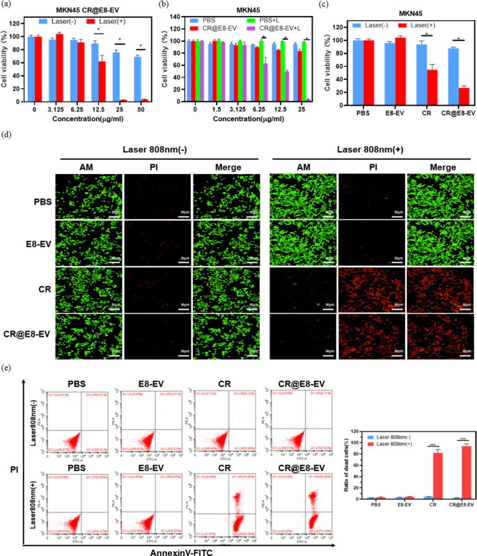
Impressed by the good photothermal efficiency of CR@E8-EVs in vitro, we then evaluated the cytotoxicity and the flexibility of photothermal ablation of CR@E8-EVs on MKN45 cells. First, the inherent photo-toxicity of CR@E8-EVs was decided by commonplace CCK8 (cell counting kit-8) viability evaluation. MKN45 cells had been incubated with CR@E8-EVs beneath numerous concentrations (24 h). As depicted inside Fig. 3a, no cytotoxicity of CR@E8-EVs to cells was noticed even with the incubation focus as much as 50 µg/mL with out irradiation, illustrating the wonderful biocompatibility. Quite the opposite, beneath 808 nm laser irradiation (0.8 W cm − 2, 5 min), CR@E8-EVs confirmed remarkably photothermal cytotoxicity to cell viability which was dramatically diminished as focus reached 25 µg/mL, at which cells had been virtually utterly eradicated (Fig. 3a). Due to this fact, CR@E8-EVs exhibited high-efficient photothermal impact in cells. However for the management therapies as proven in Fig. 3b, no apparent poisonous results for PBS, PBS plus laser and CR@E8-EVs had been noticed beneath irradiation free situation, whereas exceptional discount of cell viability was found for CR@E8-EVs plus laser. Alternatively, as proven in Fig. 3c, cells had been negligibly affected within the 4 teams with out irradiation. Nonetheless, the cell viabilities within the teams of free CR and CR@E8-EVs with laser-irradiation had been considerably decreased. Furthermore, cell viability that acquired therapy with CR@E8-EVs was decrease than that of free CR, indicating that the CR dye functionalized with engineered EVs was elevated within the uptake of cultures resulting from excessive tumor concentrating on means in contrast with free CR.
To additional visualize the photothermal ablation functionality of CR@E8-EVs, we subsequently performed Calcein-AM and PI (propidium iodide) staining of MKN45 cells by confocal scanning microscopy (Fig. 3d). For the unirradiated teams of PBS, E8-EVs, CR and CR@E8-EVs, the vast majority of cells confirmed widespread inexperienced fluorescence, implying no darkish cytotoxicity of CRs. In distinction, when irradiating the cells with a 808 nm laser for five min, the free CR and CR@E8-EVs induced apparent cell dying as proven with the purple fluorescence, validating the excessive PTT effectivity for CRs.
Moreover, the stream cytometry evaluation by Annexin V-FITC (fluoresceine isothiocyanate)/PI (propidium iodide) double staining was used to detect pro-apoptotic impact for CR dye with or with out irradiation. The leads to Fig. 3e indicated that negligible apoptosis and necrosis had been induced in all teams with out irradiation, suggesting the low toxicity of the CR dye. Whereas the teams of CR plus laser and CR@E8-EVs plus laser confirmed obvious cell apoptosis of MKN45 cells. Compared, the apoptotic charge of CR@E8-EVs plus laser was barely increased than the free CR plus laser (Fig. 3e). These outcomes had been in line with that of CCK8, additional confirming the PTT capability of CR@E8-EVs.
(a) The in vitro PTT cytotoxicity of CR@E8-EVs in MKN 45 cultures with/with out 0.8 W/cm2 808 nm laser irradiation for 10 min. Cell viability was recognized by CCK8 evaluation. (b) Cell viability of CR and PBS in opposition to MKN 45 cells and (c) relative viabilities for MKN 45 cells post-PBS (100 µL), E8-EVs (100 µL), CR (25 µg/mL, 100 µL) and CR@E8-EVs (25 µg/mL based mostly on CR, 100 µL) remedy within the presence or absence of laser. (d) Fluorescence imaging for stay/useless MKN45 cells (inexperienced/purple) with Calcein-AM and PI staining following a number of therapies. NIR gentle irradiation (808 nm, 0.8 W cm− 2, 5 min) was carried out as soon as cells had been positioned into incubation with numerous therapy for 12 h (equal to 25 µg/mL CR). Scale bars, 50 μm. (e) Apoptosis and necrosis assessments by stream cytometry in MKN 45 cells after completely different therapies. Laser irradiation (808 nm, 0.8 W cm− 2, 5 min) was carried out after cells had been incubated with numerous medicine for 12 h (PBS, 100 µL; E8-EVs, 100 µL; CR, 25 µg/mL, 100 µL and CR@E8-EVs, 25 µg/mL based mostly on CR, 100 µL)
In vivo concentrating on means of E8-EVs
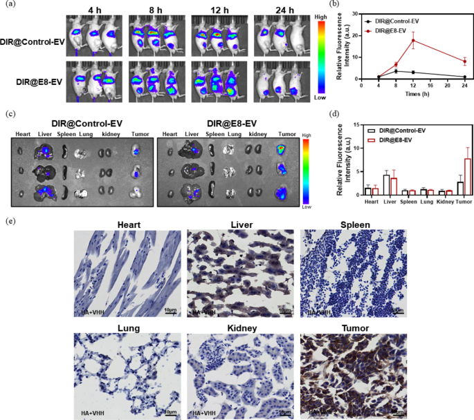
After proved the in vitro photothermal exercise of CR@E8-EVs, we subsequent investigated the in vivo concentrating on means of E8-EVs through the use of the subcutaneous MKN45 xenograft tumor mannequin. To visualise the in vivo efficiency of E8-EVs, a commercially accessible distinction agent, DIR, was harnessed to label E8-EVs (termed DIR@E8-EVs). DIR is a helpful agent in stay imaging or biomaterial tracing as a result of the emitted infrared gentle from DIR can effectively cross by cells and tissues with low background fluorescence within the infrared gentle vary. The biodistribution and accumulation had been continually tracked by an in vivo imaging system (IVIS) at pre-set timepoints after injection of DIR@E8-EVs and DIR@Con-EVs to the mice bearing MKN45 tumors (corresponding DIR dose: 1 mg/kg). As depicted in Fig. 4a, tumor fluorescence was clearly detected throughout 4 to eight h post-injection for each teams of DIR@E8-EVs and DIR@Con-EVs. However the DIR@E8-EVs exhibited a lot increased indicators than that of DIR@Con-EVs because of the lively tumor-targeting means, and the depth was elevated with time, reaching peak at 12 h for each teams. Thereafter, the imaging indicators at tumor websites had been decreased from 12 to 24 h post-administration. Nonetheless, fluorescence indicators inside tumors handled with DIR@E8-EVs had been nonetheless clear and powerful even after 24 h as in contrast with DIR@Con-EVs, which had been undetectable within the tumors after 24 h, and solely seen weak indicators within the livers (Fig. 4b). Ex vivo imaging of dissected tumors and essential organs was additional performed to verify the EVs distribution. Sign depth of DIR@E8-EVs inside tumors was a lot increased compared to DIR@Con-EVs. The sign within the management organs was not noticed besides livers (Fig. 4c). To confirm the specificity of E8-EVs in opposition to CDH17-positive tumors, one other CDH17-negative 4T1 tumor mannequin was additional used to check the imaging means of E8-EVs (Determine S5). The outcomes indicated that E8-EVs exhibited comparable tumor imaging means to control-EVs no matter in vivo imaging or ex vivo imaging in 4T1 tumor-bearing mice, implying that E8-EVs might solely exert the environment friendly payload supply in CDH17-overexpressing tumors. Additional immunohistological evaluation to important management organs and tumors within the DIR@E8-EVs group implied that no nanobody optimistic staining was noticed in essentially the most of management organs besides liver tissues, which displayed weak nanobody staining resulting from non-specific phagocytosis of reticuloendothelial system; sturdy nanobody staining was recognized within the tumors, indicating that DIR@E8-EVs might successfully focused CDH17-positive tumors (Fig. 4e). All these outcomes reveal that the E8-EVs is able to delivering CR dye for in vivo tumor concentrating on PTT.
(a) Entire physique optical imaging and (b) quantification evaluation of MKN45 tumor bearing mice at completely different postinjection timepoints. (c) Ex vivo optical imaging and (d) quantification evaluation of main organs dissected from in vivo imaged mice. (e) E8-EVs tissue distribution after 24 h injection. Scale bars, 10 μm, n = 3
In vivo photoacoustic imaging means of CR@E8-EVs
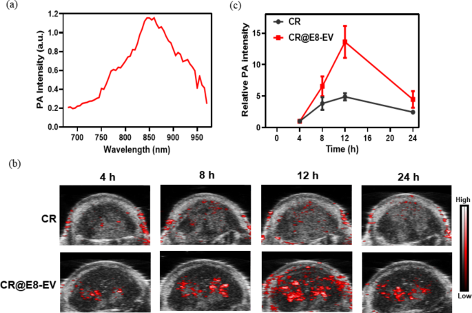
PA imaging property because the symbiotic operate for photothermal brokers, can reveal the profile of tumor tissues with excessive spatial decision [38]. Croconaine dyes have been reported for environment friendly PAI functions with ultrahigh chemical-, thermal- and photo-stability [19, 39,40,41,42]. Due to this fact, we additional evaluated the PAI property of the CR@E8-EVs on the MKN45 tumor-bearing mice. The PA photographs and depth had been monitored at diversified post-injection occasions of CR@E8-EVs; concurrently, the free CR was additionally performed as a management. The PA spectrum of CR@E8-EVs exhibited a broad band (700–950 nm) overlaying NIR-I area with a most at about 850 nm (Fig. 5a). Therefore, the in vivo PA imaging was performed at 850 nm. As depicted in Fig. 5b, PA indicators in each free CR and CR@E8-EVs turned clearly detectable at 4 h after injection, presumably because of enhanced permeability and retention (EPR) impact. Alerts without spending a dime CR from the tumor websites had been elevated barely over time and backed to the background stage at 24 h, whereas the PA indicators of CR@E8-EVs offered vital enhance because of the lively tumor-targeting means; the indicators peaked at 12 h post-injection after which decreased, implicating that 12 h post-injection was the optimum timepoint for PAI and tumor PTT therapy (Fig. 5b, c).
Imaging-driven photothermal therapy in vivo
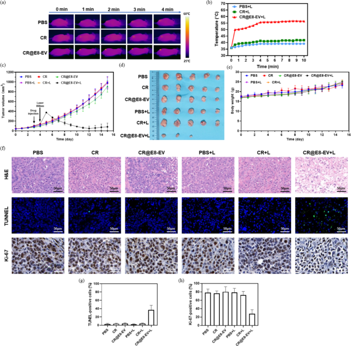
Inspired by the wonderful in vivo photothermal impact, very good tumor concentrating on efficiency and PA imaging means of CR@E8-EVs, we subsequent carried out the photothermal therapy in vivo with the MKN45 tumor xenograft mannequin and an 808 nm laser irradiation. Tumor-bearing nude mice had been stochastically separated inside six cohorts (n = 5): (1) PBS, (2) PBS plus laser, (3) CR, (4) free CR plus laser. (5) CR@E8-EVs and (6) CR@E8-EVs plus laser. For PBS, CR and CR@E8-EVs teams, 100 µL PBS, free CR and CR@E8-EVs (50 µg/mL based mostly on CR) had been injected inside tumor-bearing mice by tail vein with out laser irradiation after tumor volumes approached roughly 100 mm3. For PBS plus laser, CR plus laser and CR@E8-EVs plus laser teams, the tumors in these teams had been repeatedly irradiated with 808 nm laser (0.8 m W/cm2) for 10 min after similar dose injection. Right here, the irradiation was administered 12 h after injection in keeping with the outcomes of NIR/PA imaging, at which era level the maximal tumor accumulation was noticed. IR thermal imaging of three irradiation teams (PBS, free CR and CR@E8-EVs) was recorded by monitoring real-time temperature adjustments in tumors beneath laser irradiation (Fig. 6a,b). As illustrated in IR thermal photographs, tumor temperature for management teams handled with solely PBS and free CR demonstrated a slight enhance from 37 to 41 °C after the laser irradiation, implying that steady 808 nm laser irradiation with 0.8 W cm− 2 has negligible heat-causing impact and that free CR couldn’t successfully accumulate into tumor tissues, which is incapable of inducing apparent temperature elevation. Quite the opposite, the tumor temperature for the CR@E8-EVs plus laser group on the irradiated space quickly elevated to over 50 °C inside 40s, which was adequate to inhibit tumor progress successfully. These information additional point out that CR@E8-EVs is an efficient PTA with good penetration and excessive photothermal conversion.
To quantitatively assess the photothermal ablation efficacy of every group, tumor dimensions and mouse physique weight had been repeatedly monitored day by day post-therapy. It was discovered that the tumors grew quickly with none inhibitory results within the management teams (PBS and PBS plus laser) through the therapy interval, which indicated that 808 nm laser irradiation alone can’t inhibit tumor progress. In the meantime, the tumor progress within the free CR and CR plus laser teams was just like PBS (Fig. 6c), illustrating that CR itself possesses no concentrating on means to most cancers in vivo and can’t lead to temperature elevation after irradiation. The tumor progress within the mice acquired the therapy with CR@E8-EVs plus laser was considerably suppressed throughout the entire course of, indicating that E8-EVs might successfully ship the CR dye to tumor tissues in order that collected CR dye in tumor websites might provoke highly effective PTT efficiency to manage tumor development (Fig. 6c). By the tip of the therapy process, all of the mice had been sacrificed and tumors had been photographed (Fig. 6d). The tumor photographs confirmed the identical pattern with the quantity, and the common tumor measurement within the CR@E8-EVs plus laser group was considerably diminished with a comparatively small tumor quantity and even two tumors disappeared (Fig. 6d). Photothermal remedy as a therapeutic modality cannot solely immediately ablate tumors but in addition provoke the anti-tumor immunity and reprogram tumor microenvironment by the induction of immunogenic cell dying (ICD), even induce long-term immune reminiscence, which has been validated in competent mouse tumor fashions [43,44,45,46,47], which could the principle trigger in present examine for tumor eradication in two mice the place macrophages or dendritic cells could possibly be intensely activated in our compromise tumor mannequin and phagocytosis is likely to be enhanced. Additional investigation is have to decipher the adjustments in anti-tumor immunity and tumor microenvironment in competent mouse tumor mannequin after CR-mediated PTT. These tumor inhibition findings had been in line with the outcomes of in vitro experiments, which additional verifies the wonderful PTT capability of CR@E8-EVs. Moreover, no obvious body-weight loss and medical abnormality had been noticed in all mice in these six teams over the 15-day therapy interval, suggesting that photothermal therapy has good in vivo biosafety (Fig. 6e).
To additional consider the impact of tumor inhibition and the biosafety of PTA, histopathological evaluation of tumors and main management organs (coronary heart, liver, spleen, lungs, kidneys and colons) was carried out by commonplace hematoxylin and eosin (H&E) staining after remedy. No noticeable tissue harm/abnormality and inflammatory lesion of the management organs had been recognized from all of the teams as proven in Fig. S6, demonstrating the favorable biosafety of CRs/EVs-based PTT. Moreover, H&E staining evaluation for tumor tissues revealed that CR@E8-EVs plus laser irradiation confirmed extreme cell harm with extra sparse tissues, whereas no apparent harm was noticed within the different teams (Fig. 6f). Furthermore, TUNEL evaluation for cell apoptosis evaluation unraveled that CR@E8-EVs plus laser therapy induced vital cell apoptosis in contrast with the opposite teams; and additional Ki67 staining indicated that tumor cell proliferation was considerably inhibited in CR@E8-EVs plus laser group in contrast with management teams (Fig. 6f). The entire outcomes above implicate that CR@E8-EV- based mostly PTT might exert very good anti-tumor efficiency and engineered EVs is an efficient technique for the supply of CR dyes to provide distinguished photothermal impact on tumors.
(a) Photothermal imaging and (b) tumor temperature adjustments beneath 808 nm laser irradiation. (c) Tumor progress curves following numerous therapies. (d) Tumor dissection pictures for all six teams (n = 5) at day 16 post-therapy. (e) Physique weight of mice throughout therapy. (f) Histological H&E, and fluorescence TUNEL and Ki67 staining for tumor tissues on the finish of the therapy (scale bars, 50 μm). (g) Quantification of TUNEL-positive cells. (h) Quantification of Ki67 optimistic cells


