

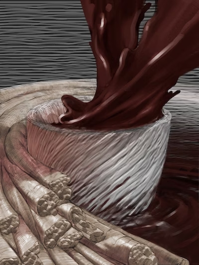
This illustration reveals a 3D printed coronary heart ventricle engineered with fiber-infused ink. Credit score: Harvard SEAS
By Kat J. McAlpine / SEAS Communications
During the last decade, advances in 3D printing have unlocked new potentialities for bioengineers to construct coronary heart tissues and buildings. Their targets embrace creating higher in vitro platforms for locating new therapeutics for coronary heart illness, the main reason behind dying in the USA, accountable for about one in each 5 deaths nationally, and utilizing 3D-printed cardiac tissues to judge which remedies would possibly work finest in particular person sufferers. A extra distant purpose is to manufacture implantable tissues that may heal or exchange defective or diseased buildings inside a affected person’s coronary heart.
In a paper printed in Nature Supplies, researchers from Harvard John A. Paulson Faculty of Engineering and Utilized Sciences (SEAS) and the Wyss Institute for Biologically Impressed Engineering at Harvard College report the event of a brand new hydrogel ink infused with gelatin fibers that permits 3D printing of a useful coronary heart ventricle that mimics beating like a human coronary heart. They found the fiber-infused gel (FIG) ink permits coronary heart muscle cells printed within the form of a ventricle to align and beat in coordination like a human coronary heart chamber.
“Individuals have been making an attempt to copy organ buildings and capabilities to check drug security and efficacy as a means of predicting what would possibly occur within the medical setting,” says Suji Choi, analysis affiliate at SEAS and first writer on the paper. However till now, 3D printing methods alone haven’t been capable of obtain physiologically-relevant alignment of cardiomyocytes, the cells accountable for transmitting electrical indicators in a coordinated trend to contract coronary heart muscle.
“We began this venture to deal with a number of the inadequacies in 3D printing of organic tissues.”
– Kevin “Package” Parker
The innovation lies within the addition of fibers inside a printable ink. “FIG ink is able to flowing by way of the printing nozzle however, as soon as the construction is printed, it maintains its 3D form,” says Choi. “Due to these properties, I discovered it’s attainable to print a ventricle-like construction and different complicated 3D shapes with out utilizing further assist supplies or scaffolds.”
This video reveals the spontaneous beating of a 3D-printed coronary heart muscle. Credit score: Harvard SEAS.
To create the FIG ink, Choi leveraged a rotary jet spinning approach developed within the lab of Kevin “Package” Parker, Ph.D. that fabricates microfiber supplies utilizing an strategy just like the best way cotton sweet is spun. Postdoctoral researcher and Wyss Lumineer Luke MacQueen, a co-author on the paper, proposed the concept that fibers created by the rotary jet spinning approach might be added to an ink and 3D printed. Parker is a Wyss Affiliate College member and the Tarr Household Professor of Bioengineering and Utilized Physics at SEAS.
“When Luke developed this idea, the imaginative and prescient was to broaden the vary of spatial scales that might be printed with 3D printers by dropping the underside out of the decrease limits, taking it right down to the nanometer scale,” Parker says. “The benefit of manufacturing the fibers with rotary jet spinning slightly than electrospinning” – a extra typical methodology for producing ultrathin fibers – “is that we will use proteins that may in any other case be degraded by {the electrical} fields in electrospinning.”
Utilizing the rotary jet to spin gelatin fibers, Choi produced a sheet of fabric with an identical look to cotton. Subsequent, she used sonification – sound waves – to interrupt that sheet into fibers about 80 to 100 micrometers lengthy and about 5 to 10 micrometers in diameter. Then, she dispersed these fibers right into a hydrogel ink.
“This idea is broadly relevant – we will use our fiber-spinning approach to reliably produce fibers within the lengths and shapes we wish.”
– Suji Choi
Essentially the most troublesome facet was troubleshooting the specified ratio between fibers and hydrogel within the ink to take care of fiber alignment and the general integrity of the 3D-printed construction.
As Choi printed 2D and 3D buildings utilizing FIG ink, the cardiomyocytes lined up in tandem with the course of the fibers contained in the ink. By controlling the printing course, Choi may subsequently management how the guts muscle cells would align.
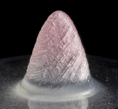
The tissue-engineered 3D ventricle mannequin. Credit score: Harvard SEAS
When she utilized electrical stimulation to 3D-printed buildings made with FIG ink, she discovered it triggered a coordinated wave of contractions in alignment with the course of these fibers. In a ventricle-shaped construction, “it was very thrilling to see the chamber truly pumping in an identical strategy to how actual coronary heart ventricles pump,” Choi says.
As she experimented with extra printing instructions and ink formulation, she discovered she may generate even stronger contractions inside ventricle-like shapes.
“In comparison with the actual coronary heart, our ventricle mannequin is simplified and miniaturized,” she says. The group is now working towards constructing extra life-like coronary heart tissues with thicker muscle partitions that may pump fluid extra strongly. Regardless of not being as robust as actual coronary heart tissue, the 3D-printed ventricle may pump 5-20 instances extra fluid quantity than earlier 3D-printed coronary heart chambers.
The group says the approach may also be used to construct coronary heart valves, dual-chambered miniature hearts, and extra.
“FIGs are however one device we now have developed for additive manufacturing,” Parker says. “We’ve different strategies in growth as we proceed our quest to construct human tissues for regenerative therapeutics. The purpose is to not be device pushed – we’re device agnostic in our seek for a greater strategy to construct biology.”
Extra authors embrace Keel Yong Lee, Sean L. Kim, Huibin Chang, John F. Zimmerman, Qianru Jin, Michael M. Peters, Herdeline Ann M. Ardoña, Xujie Liu, Ann-Caroline Heiler, Rudy Gabardi, Collin Richardson, William T. Pu, and Andreas Bausch.
This work was sponsored by SEAS; the Nationwide Science Basis by way of the Harvard College Supplies Analysis Science and Engineering Heart (DMR-1420570, DMR-2011754); the Nationwide Institutes of Well being and Nationwide Heart for Advancing Translational Sciences (UH3HL141798, 225 UG3TR003279); the Harvard College Heart for Nanoscale Programs (CNS), a member of the Nationwide Nanotechnology Coordinated Infrastructure Community (NNCI) which is supported by the Nationwide Science Basis (ECCS-2025158, S10OD023519); and the American Chemical Society’s Irving S. Sigal Postdoctoral Fellowships.
![]()
Wyss Institute
makes use of Nature’s design ideas to develop bioinspired supplies and units that may rework medication and create a extra sustainable world.
![]()
Wyss Institute
makes use of Nature’s design ideas to develop bioinspired supplies and units that may rework medication and create a extra sustainable world.
