Characterization and LDI-MS efficiency of IVONS:Sm
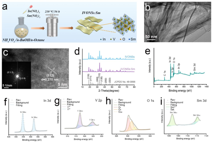
As depicted in Fig. 1a, the preparation of IVONSs:Sm was carried out utilizing the microemulsion-mediated solvothermal technique. Transmission electron microscopy (TEM) evaluation revealed that the as-synthesized IVONSs:Sm have been of translucence and corrugation, indicative of a sheet-like nanostructure which was conducive to the adsorption of LMW molecules (Fig. 1b). A lattice spacing of 0.270 nm in excessive decision TEM (HRTEM) picture was listed to (112) airplane of orthorhombic InVO4 (Fig. 1c). Moreover, the chosen space electron diffraction (SAED) sample confirmed the excessive diploma of crystallinity of IVONSs:Sm (inset, Fig. 1c). Elemental evaluation utilizing power dispersive spectrum (EDS) and EDS mapping additional verified the presence and uniform distribution of In, V, O, and Sm components in IVONSs:Sm, with semiquantitative outcomes intently aligning with the theoretical compositions (Determine S1a, b). Determine 1d exhibited the powder X-ray diffraction (XRD) patterns of IVONSs:Sm, the sharp diffraction peaks additionally revealed the great crystallinity of IVONSs:Sm and matched nicely with the usual orthorhombic-phase InVO4 (JCPDS No. 48–0898). It’s noteworthy that Sm doping had no impact on the attribute diffraction sample of InVO4, and there was no impurity peak involved with In2O3, V2O5 or different species. Past that, survey X-ray photoelectron spectroscopy (XPS) evaluation confirmed the weather of In, V, O, and Sm with out impurities (Fig. 1e). Moreover, high-resolution XPS spectra of In 3d, V 2p, O 1s, and Sm 3d offered detailed insights into the chemical composition and bonding states of the IVONSs:Sm (Fig. 1f ~ i). The high-resolved In 3d spectrum was characterised by an In 3d5/2 peak at 444.3 eV and an In 3d3/2 peak at 452.1 eV (Fig. 1f). For the V 2p spectrum, the fitted peaks at 516.9 eV and 525.0 eV have been related to V 2p3/2 and V 2p1/2 of V5+, respectively. Whereas peaks positioned at 515.2 eV and 523.3 eV have been assigned to V 2p orbitals of V4+, indicating V5+ may receive electrons from close by oxygen vacancies (Fig. 1g). As anticipated, O 1s spectrum revealed three deconvoluted peaks at 530.3, 532.1 and 533.6 eV, representing lattice oxygen (OL), emptiness oxygen (OV) and chemisorbed oxygen (OOH), respectively (Fig. 1h). Within the elements of Sm 3d peak, two peaks fitted at 1083.5 eV and 1110.9 eV have been corresponded to Sm 3d5/2 and Sm 3d3/2 orbitals, respectively (Fig. 1i). Collectively, these characterization outcomes offered proof of the profitable synthesis of IVONSs:Sm.
(a) Schematic illustration of IVONSs:Sm preparation based mostly on the microemulsion-mediated solvothermal technique. (b) TEM picture, (c) HRTEM picture, and SAED sample (inset) of IVONSs:Sm. (d) XRD spectra of IVONSs:Sm and pure IVONSs. XPS evaluation of IVONSs:Sm: (e) Survey spectrum, (f) In 3d, (g) V 2p, (h) O 1s, and (i) Sm 3d
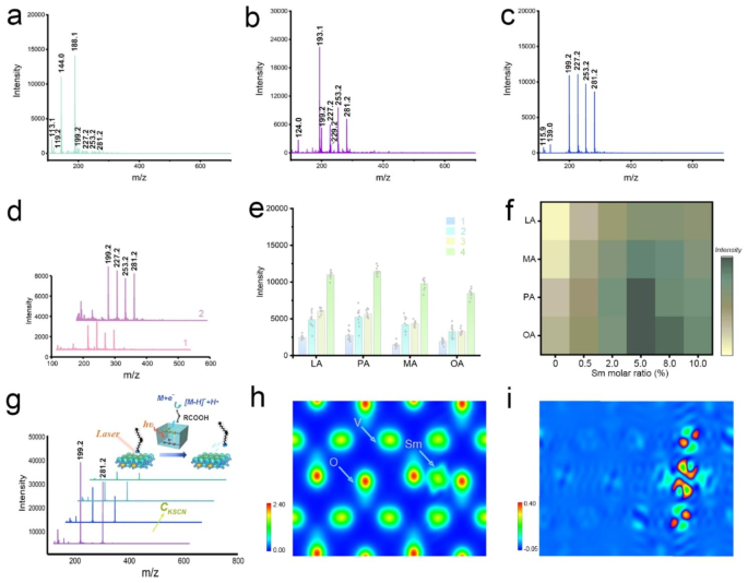
To be able to consider the efficiency of IVONSs:Sm as a nano-matrix for LDI-MS evaluation, the 4 commonest fatty acids in organisms – lauric acid (LA), myristic acid (MA), palmitoleic acid (PA), and oleic acid (OA) – have been chosen as mannequin analytes and analyzed with IVONSs:Sm, in addition to conventional natural matrices (CHCA and 9-AA) in each positive- and negative-ion modes. When CHCA was utilized within the positive-ion mode, the cationic adducts and fragments of CHCA have been discovered to be predominant, inhibiting the MS alerts of the 4 analyte ions, together with [M + H]+ and [M + Na]+, underneath these background alerts (Determine S2a, Desk S2). Moreover, Determine S2b additionally revealed a suppressive MS sign of the 4 fatty acids, indicating the inapplicability of 9-AA in positive-ion MALDI-MS evaluation. As proven in Determine S2c, a number of positive-ion alerts of fatty acids may very well be detected with much less interference peak when IVONSs:Sm was used as a nano-matrix. Nonetheless, it’s noteworthy that accompanying the quasi-molecular ion peaks of fatty acids have been the alkali and double alkali adduct ions of the analytes (Desk S2), rendering the mass spectrum in positive-ion mode notably difficult to interpret. These outcomes suggest that it might not be a superb choice to research fatty acids within the positive-ion mode. Alternatively, Fig. 2a and b demonstrated the MS spectra of the 4 fatty acids in negative-ion mode utilizing CHCA and 9-AA as matrices. For CHCA, the matrix-related ions ([M − CO2 − H]− at m/z 144.0 and [M − H] − at m/z 188.1) dominated, however suppressive analyte alerts have been noticed. As well as, improved MS sign depth and decrease lacking share of fatty acids have been noticed for the 9-AA matrix (Desk S2). However, a better depth of intrinsic matrix-related ion ([M-H]− at m/z 193.1) nonetheless suppressed the MS sign of fatty acids. In distinction, an interference-free MS spectrum with deprotonated [M − H]− ions of LA, MA, PA, and OA at m/z 199.2, 227.2, 253.2, and 281.2 was obtained within the vary of m/z 150–600 with IVONSs:Sm because the nano-matrix (Fig. 2c), implying that IVONSs:Sm have been extra relevant in facilitating destructive ionization for fatty acids. As a management, the efficiency of the three matrices with out analytes was additionally evaluated (Determine S3). The outcomes indicated that backgrounds with a number of intrinsic matrix-related peaks have been noticed for these utilizing CHCA and 9-AA as matrices in numerous ionization modes, whereas much less background interference for IVONSs:Sm ranged from m/z 150 to m/z 600, particularly in negative-ion mode, additional demonstrating the potential of IVONSs:Sm as a secure nano-matrix for LDI-MS evaluation.
MS spectra of 4 fatty acids analyzed through the use of (a) CHCA, (b) 9-AA, and (c) IVONSs:Sm in negative-ion mode, respectively. (d) MS spectra of 4 fatty acids analyzed through the use of graphene (1) and CeO2 (2) in negative-ion mode. (e) MS depth comparability amongst 4 fatty acids utilizing totally different nano-matrices: graphene (1), CeO2 (2), IVONSs (3), and IVONSs:Sm (4). (f) Optimization of MS sign depth versus the molar ratio of Sm doping quantity for IVONS:Sm. (g) IVONSs:Sm-assisted LDI-MS spectra of LA and OA mixtures with totally different concentrations of KSCN in negative-ion mode, and the corresponding schematic diagram of the negative-ion LDI mechanism based mostly on IVONSs:Sm (inset). (h) Electron density profile and (i) differential electron density profile of IVONSs:Sm
Moreover, the research evaluated the efficiency of LDI-MS utilizing IVONSs:Sm in distinction to beforehand reported nano-matrices equivalent to graphene and metallic oxide (e.g., cerium oxide (CeO2)), which have been acknowledged for his or her superiority in SALDI-MS evaluation of LWM compounds (Determine S4a, b) [34, 37]. Determine 2c, d revealed that MS alerts of the 4 fatty acids may very well be distinctly detectable as [M − H]− utilizing the three nano-matrices. Nonetheless, the upper analyte alerts for IVONSs:Sm steered its superiority over graphene and CeO2 in detection sensitivity (Fig. 2e), which may very well be ascribed to a synergistic impact of stronger UV absorption, increased photothermal functionality of IVONSs:Sm, and probably efficient interactions between IVONSs:Sm and floor LMW compounds. As proven in Determine S4c, UV-vis absorption spectra of the three nano-matrices with the identical dispersing focus have been investigated. A stronger absorption of IVONSs:Sm than different nano-matrices on the operational wavelength (355 nm) made it doable to be utilized as a benign receptor of laser power within the LDI-MS course of. Moreover, the infrared thermographic photographs and photothermal heating curves of the three nano-matrices indicated that IVONSs:Sm had extra advantageous photothermal conversion in comparison with the opposite two standard nano-matrices, which made it efficient to switch the absorbed laser power and improve the ionization of analytes (Determine S5). With this in thoughts, it may very well be an optimum alternative to research LMW compounds with IVONSs:Sm as a nano-matrix. Moreover, Fig. 2e and Determine S6 steered that the incorporation of Sm3+ into IVONSs may very well be synergistically contributive to the destructive ionization course of. Considerably, the MS spectrum with the identical attribute peaks of the 4 fatty acids however inadequate MS intensities was obtained through the use of IVONSs with out samarium doping. This end result demonstrated the superior feasibility of IVONSs:Sm over pure IVONSs within the evaluation of fatty acids. Accordingly, the optimum dopant quantity was investigated, and Fig. 2f indicated that the MS sign intensities of the 4 fatty acids reached their most at 5.0 mol % samarium, suggesting an optimum samarium molar ratio for IVONSs:Sm.
Mechanistic foundation of IVONSs:Sm-assisted LDI-MS
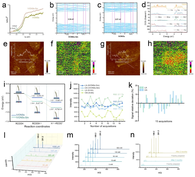
Whereas the exact LDI mechanism stays to be conclusively established, quite a few research have offered proof supporting the photoexcitation and digital transition mechanism induced by laser within the ionization course of [38]. Therefore, the enhancement of MS alerts with IVONSs:Sm may be elucidated by the digital transition mechanism for the destructive ionization of LMW molecules. As depicted in Fig. 2g (inset), the enter laser power may very well be transduced by technology of electron-hole pairs in IVONSs:Sm, the place the excited electrons may very well be emitted from IVONSs:Sm and subsequently work together with LMW molecules, facilitating the destructive ionization of the analyte [M − H]−. To validate this mechanistic foundation, potassium thiocyanate was employed as a hole-scavenger to analyze the correlation between the technology of electron-hole pairs and the LDI strategy of LMW molecules [38]. Determine 2g demonstrated reducing deprotonated ion peaks of of LA (m/z 199.2) and OA (m/z 281.2) with a rise in KSCN focus (0, 5, 10, 20 µmol/mL), exhibiting a definite suppression of the MS sign of the 2 fatty acids within the presence of upper KSCN focus, thus experimentally supporting the digital transition mechanism in Fig. 2g [39]. Consequently, the digital construction and optoelectronic properties of IVONSs:Sm ought to be examined. Determine 2h, i and Determine S7 offered the electron density evaluation of IVONSs and IVONSs:Sm on the crystal face (100). Compared to pure IVONSs (Determine S7), the distinct electron cloud overlapping in IVONSs:Sm confirmed the coexistence state of electrovalent bond and covalent bond in samarium-oxygen polarity linkages (Fig. 2h). Accordingly, the differential electron density diagram indicated that the electron density of O2- adjoining to Sm3+ elevated (Fig. 2i). Thus, the octahedron fashioned by Sm3+-O2- bond induced the enhancement of {the electrical} dipole second within the lattice, which was useful for the diffusion of the cost provider. Alternatively, Determine S8a depicted the diffuse reflectance ultraviolet-visible spectra of IVONSs:Sm and pure IVONSs. Notably, the sturdy absorption bands starting from 200 to 400 nm have been primarily attributed to the cost switch impact between V5+–O2- and f-f transitions of Sm3+ (Determine S8b) [40, 41], implying that IVONSs:Sm predominantly absorbed laser energy (e.g., 355 nm Nd:YAG laser) within the MALDI-MS instrument, thus permitting for the next ionization course of. In response to the Tauc equation, the corresponding band gaps of IVONSs:Sm and pure IVONSs based mostly on the optical absorption have been calculated to be 2.19 and a couple of.28 eV, respectively (Fig. 3a). The decrease band hole of IVONSs:Sm than pure IVONSs was additional supported by density purposeful principle (DFT) simulation (Fig. 3b, c), the place the DFT-derived band hole of pure IVONSs aligned with the experimental knowledge. For IVONSs:Sm, the direct band hole of IVONSs:Sm was barely smaller than the experimental worth, as DFT simulation tends to underestimate the band hole [42]. The density of states (DOS) outcomes of IVONSs indicated that the conduction band was predominantly composed of V 3d states (Determine S9a), whereas the V 3d states hybridized with the Sm 4f states, forming the conduction band of IVONSs:Sm (Fig. 3d). This steered that the Sm 4f states would shift the conduction band in the direction of the decrease power, thus decreasing the band hole for IVONSs:Sm. The experimental and calculation outcomes constantly demonstrated the lower within the IVONSs band hole with Sm ion dopant. Importantly, the decrease band hole of IVONSs:Sm promoted the digital transition and cost provider technology, which was conducive to the destructive ionization of analytes as per the mechanism illustrated above.
(a) Tauc plots of IVONSs:Sm and IVONSs. Vitality band construction of (b) IVONSs and (c) IVONSs:Sm. (d) Calculated DOS of IVONSs:Sm. (e) AFM and (f) SPV photographs of IVONSs, and (g) AFM and (h) SPV photographs of IVONSs:Sm. (i) Calculated Gibbs free-energy diagram for the adsorption and dissociation strategy of fatty acid over IVONSs:Sm and IVONSs surfaces, respectively. (j) Reproducibility of MS sign intensities for LA and OA utilizing totally different matrices. (okay) Sign relative deviation of MS sign intensities utilizing IVONSs:Sm. (l) IVONSs:Sm-assisted LDI-MS spectra of various concentrations of LA and OA in negative-ion mode. (m) IVONSs:Sm-assisted LDI-MS spectra of LA and OA (500 µM) in numerous concentrations of NaCl in negative-ion mode. (n) IVONSs:Sm-assisted LDI-MS spectra of a clean pattern (inexperienced traces) and a combination of LA and OA (yellow traces) with freshly ready IVONSs:Sm and IVONSs:Sm after two months of preservation
As well as, Fig. 3e ~ h depicted atomic power microscope (AFM) and floor photovoltage (SPV) photographs of the 2 vanadate nanosheets, offering a spatial visualization of the technology and transport of cost carriers on the nanoscale. The AFM photographs of IVONSs:Sm and pure IVONSs, with comparable lateral dimensions and thickness of roughly 5 nm, have been offered in Fig. 3e and g. Nonetheless, the corresponding SPV photographs, obtained by subtracting the potentials underneath darkish circumstances, revealed that IVONSs:Sm exhibited a extra destructive light-induced potential change of roughly 30 mV in comparison with pure IVONSs. This indicated a better accumulation of electrons and larger mobility of cost carriers from the majority part to the floor of IVONSs:Sm, which facilitated power switch from the nanomaterial to LMW molecules, consequently growing the effectivity of destructive ionization in LDI-MS. To achieve perception into the function of the nanomaterial IVONSs:Sm within the LDI course of, we additional investigated the thermodynamic profile of adsorption and dissociation of analytes on the 2 vanadate nanosheets utilizing DFT simulations. As a consultant LMW molecule, the electrostatic potential (ESP) of OA was initially calculated based mostly on the bottom state electron density (Determine S9b). The carboxyl group was discovered to be favorable for OA (RCOOH) to be adsorbed onto the vanadate nanosheets (RCOOH*) because of its most ESP. Consequently, the acidic hydrogen within the carboxyl terminal tended to switch from OA to the floor lattice oxygen, liberating the destructive ion of OA (RCOO−). Determine 3i demonstrated that the Gibbs free energies of every step have been decreased on IVONSs:Sm relative to pure IVONSs, indicating that the LDI course of on the surfaces of IVONSs:Sm was thermodynamically favorable. Taking all the above into consideration, the improved efficiency of IVONSs:Sm in nanomaterial-assisted LDI may very well be elucidated as a synergistic impact of assorted components, together with optical absorption and cost provider mobility.
IVONSs:Sm-assisted LDI-MS evaluation of fatty acids
Resulting from its superior efficiency, IVONSs:Sm was decided to be a extremely efficient nano-matrix for in situ detection of LMW compounds in genuine samples, equivalent to fingerprints. A lifting course of utilizing double-sided conductive copper foil tape was carried out to retrieve fingerprints from surfaces. The distinctive potential of IVONSs:Sm in negatively ionizing LMW compounds on the copper conductive tape was additionally verified (Determine S10). To evaluate the reproducibility of IVONSs:Sm-assisted LDI-MS in negative-ion mode, the mass spectrometry sign intensities of consultant LA (saturated fatty acid) and OA (unsaturated fatty acid) from fifteen randomly chosen positions inside a single spot (diameter ~ 2.1 mm) of analytes noticed on the copper conductive tape have been collected. As proven in Fig. 3j, comparatively secure MS alerts for LA and OA have been noticed, and a low relative commonplace deviation for every acquisition confirmed the excessive shot-to-shot reproducibility of the IVONSs:Sm-assisted LDI-MS strategy (Fig. 3okay). Compared, the MS sign intensities for LA and OA confirmed important fluctuations with an RSD of ~ 20% for every acquisition (Fig. 3j), indicating unsatisfactory repeatability when using the standard natural matrix 9-AA. The nice shot-to-shot reproducibility was primarily attributed to the distinction within the homogeneity of IVONSs:Sm and 9-AA deposited on the copper conductive tape. Determine S11 demonstrated the crystallization of IVONSs:Sm and two forms of conventional natural matrices (CHCA and 9-AA) obtained in a MALDI-MS spectrometer, through which a stark distinction of matrix distribution between IVONSs:Sm and different two natural matrices may very well be witnessed. It may very well be seen that IVONSs:Sm unfold extra uniformly after the solvent evaporation, as a substitute of exhibiting the “candy spot” impact of CHCA and 9-AA on the goal floor. The nice shot-to-shot reproducibility successfully solved the variability of sign depth, offering assurance of dependable quantitative evaluation of analytes utilizing IVONSs:Sm. Fig. 3L demonstrated that the MS peaks of the 2 deprotonated fatty acids [M − H]− steadily raised with the growing analyte concentrations. The MS responses for LA at m/z 199.2 and OA at m/z 281.2 have been proportional to the concentrations of the goal analytes (Determine S13a), and the detection restrict (LOD) investigations of LA and OA corroborated the sensitivity of the IVONSs:Sm-assisted LDI-MS strategy (8.2 µM for LA and 11.6 µM for OA). As proven in Determine S12, a quantitative evaluation of residual LA and OA in actual fingerprint samples was additional carried out utilizing high-performance liquid chromatography-electrospray ionization mass spectrometry (HPLC-ESI-MS). The residual portions of LA and OA in a fingerprint pattern have been decided to be 0.36 and a couple of.72 µg, respectively. Taking the LODs and added quantity (1 µL) of fatty acid commonplace options into consideration, the proposed IVONSs:Sm-assisted LDI-MS strategy was able to detecting 1.6 × 10− 3 µg of LA and three.3 × 10− 3 µg of OA in actual samples, which have been considerably decrease than these detected by HPLC-ESI-MS. Subsequently, the quantitative outcomes supported that IVONSs:Sm-assisted LDI-MS may fulfill the analytical demand of fatty acid ranges in fingerprints. Given {that a} excessive salt focus may suppress the ionization strategy of the analyte, the salt tolerance was evaluated by including totally different concentrations of NaCl (0 ~ 500 mM) in IVONSs:Sm-assisted LDI-MS detection of two consultant fatty acids. Determine 3m confirmed that the addition of 10–500 mM NaCl barely decreased the sign intensities of LA and OA, confirming a superb tolerance of the nano-matrix IVONSs:Sm. Moreover, it was famous that the attribute MS peaks of the LA and OA combination utilizing IVONSs:Sm with a two-month storage matched these utilizing freshly ready IVONSs:Sm (Fig. 3n), and there was insignificant divergence within the sample and depth of the MS peaks after a number of laser pictures (Determine S13b, c). Therefore, the steadiness of IVONSs:Sm, together with the photostability of analytes throughout IVONSs:Sm-assisted LDI-MS evaluation, may very well be experimentally ascertained.
IVONSs:Sm-assisted LDI-MS imaging of fingerprints
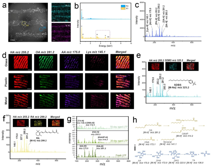
Based mostly on the aforementioned research, the applicability of IVONSs:Sm for detecting LMW compounds adhered to fingerprints was preliminarily investigated. IVONSs:Sm have been dispersed in bromoethane with a low boiling level after which sprayed on the fingerprint part by an atomizer. Following the deposition of IVONSs:Sm, the fingerprint was lifted utilizing copper conductive tape. Scanning electron microscopy (SEM) photographs and outcomes from EDS evaluation confirmed the focus of IVONSs:Sm on the ridges of the fingerprint after the disperse medium evaporation (Fig. 4a, b). To gather IVONSs:Sm-assisted LDI-MS spectra of the fingerprints, the copper conductive tape containing the extracted fingerprint was affixed to an indium tin oxide-coated glass slide for IVONSs:Sm-assisted LDI-MS evaluation. Determine S14a offered the optimistic MS profile obtained from the extracted fingerprint, which confirmed the MS spectrum of an endogenous combination containing PA, hexadecanoic acid (HA), OA, and stearic acid (SA). The abundance of MS peaks confirmed that the chemical info of the fingerprint has been transferred onto the substrate of the copper conductive tape. Other than the fatty acids, different ion peaks originated from different endogenous compounds have been additionally recorded within the vary m/z 100–700. For instance, the peaks detected at m/z 147.1 and 177.1 have been assigned to the [M + H]+ ion of lysine (Lys) and ascorbic acid (AA), respectively. In distinction, the MS spectrum of the extracted fingerprint obtained in negative-ion mode exhibited a comparatively concise sign with long-term stability in excessive vacuum (Fig. 4c and Determine S14b ~ d). In contrast to the a number of peaks for one analyte underneath positive-ion mode, all of the fatty acids have been clearly detected as the one deprotonated [M − H]− ions with the help of IVONSs:Sm. Moreover, intense ions within the fingerprint have been putatively recognized by way of MALDI LIFT-TOF/TOF MS (Determine S15, S16, and Desk S3). Moreover, contemplating the coexistent lipids in fingerprints, in addition to the potential ester bond cleavage within the case of IVONSs:Sm, a managed experiment was carried out and indicated a comparatively secure MS sign of two mannequin fatty acids within the existence of two typical lipids (glycerol trimyristate and glycerol tripalmitate), which excluded the interference of lipids on subsequent fatty acid evaluation (Determine S17). All the outcomes steered the preponderance and feasibility of negative-ion IVONSs:Sm-assisted LDI-MS for spatial molecular profiling in fingerprints.
(a) SEM picture of a fingerprint sprayed with IVONSs:Sm and corresponding EDS mapping of In and V components. (b) EDS spectra of round areas (ridge and valley in a fingerprint) within the SEM picture. (c) IVONSs:Sm-assisted LDI-MS spectra of a fingerprint in negative-ion mode. (d) MS photographs of the fingerprints collected from totally different substrates in negative-ion mode. IVONSs:Sm-assisted LDI-MS evaluation for (e) SDBS and (f) RA on fingerprints. (g) IVONSs:Sm-assisted LDI-MS spectra of recent and aged fingerprints in negative-ion mode. Proposed oxidation pathway of (h) OA and (i) AA throughout fingerprint storage. Scale bars in (d)~(f) signify 1 mm
Since IVONSs:Sm allowed for the MS evaluation of chemical species contained inside the fingerprint, IVONSs:Sm-assisted LDI-MS imaging of the fingerprint may very well be generated from the spatial distribution of the MS alerts of chosen deprotonated analytes. Contemplating the spraying circumstances of IVONSs:Sm may have an effect on the loading quantity and homogeneous protection of the nano-matrix on the fingerprint, crucial experimental parameters (together with dispersing focus of IVONSs:Sm and spraying time) have been optimized. For the sake of simplicity, HA was taken because the consultant fatty acid for IVONSs:Sm-assisted LDI-MS evaluation in negative-ion mode. As proven in Determine S18a, the vanadium content material on the substrate of the copper conductive tape was decided by inductively coupled plasma mass spectrometry (ICP-MS), which implied that the loading quantity of IVONSs:Sm on the fingerprint steadily elevated with the rise of IVONSs:Sm focus (Determine S18b). As a consequence, the MS sign of HA at m/z 255.2 went up with the dispersing focus of IVONSs:Sm. Likewise, the spraying time was optimized, and the outcomes have been offered in Determine S18c. The incremental vanadium content material and the MS sign with the spraying time of IVONSs:Sm suspension additional confirmed that satisfactory IVONSs:Sm deposition was useful to the enhancement of the MS sign. Other than the analysis of spraying circumstances in mild of MS sign depth of the analyte, the efficiency of IVONSs:Sm-assisted LDI-MS imaging may additionally rely upon parameters just like the dispersing focus of IVONSs:Sm and spraying time. It may very well be discovered that the MS photographs with increased picture readability and distinction have been obtained with the growing focus of IVONSs:Sm (Determine S19a ~ c). Determine S19d ~ f indicated that the MS photographs of the fingerprint steadily blurred when the spraying time elevated to 60 s, suggesting that the extended deposition process of IVONSs:Sm resulted in an undesirable MS picture with out the unique trivia of the fingerprint sample. This phenomenon is perhaps ascribed to the issue that prolonged and steady publicity of sprayed bromoethane droplets may totally moist the fingerprint space, inflicting the delocalization and diffusion of hydrophobic fatty acids, thus the corresponding MS picture of the fingerprint was obtained with poor spatial decision. On this case, the correct spraying time enabled a well-defined MS picture in fingerprint evaluation. Based mostly on the above outcomes, the optimum parameters of dispersing focus (5 mg/mL) and spraying time (30 s) of IVONSs:Sm suspension for the matrix deposition may very well be settled.
To be able to examine the feasibility of the steered IVONSs:Sm-assisted LDI-MS technique for fingerprint evaluation, the fingerprints left on totally different materials surfaces have been analyzed by way of IVONSs:Sm-assisted LDI-MS imaging in negative-ion mode. As proven in Fig. 4d, the fingerprint morphological options may very well be clearly perceived, and the MS photographs may additionally present the spatial distribution of attribute LMW compounds detected at m/z 145.1 (Lys), 175.0 (AA), 255.2 (HA), and 281.2 (OA). Importantly, there was no particular distinction amongst these three materials surfaces in imaging high quality. These outcomes show the potential of IVONSs:Sm-assisted LDI-MS imaging to permit molecular-level fingerprint recognition on totally different materials surfaces. Determine S20 demonstrated that the imaging impact utilizing IVONSs:Sm in contrast favorably with these of graphene and CeO2. In comparison with graphene and CeO2 (Desk S4), the best sign depth and the most important variety of detectable endogenous LMW compounds in fingerprints have been obtained utilizing IVONSs:Sm (Determine S20a). As seen in Determine S20b, fingerprint morphological options and spatial distribution of two consultant LMW compounds (OA and Lys) may very well be clearly visualized, whereas comparatively decrease MS intensities of LMW compounds from the nano-matrix graphene affected the utilized efficiency of MS imaging for the fingerprint (Determine S20c). Furthermore, a decrease distinction between ridges and valleys of the fingerprint was noticed from the MS photographs utilizing the nano-matrix CeO2 (Determine S20d). This phenomenon is perhaps primarily ascribed to the low hydrophobicity of the nano-matrix CeO2 (Determine S21), which led to an uneven dispersion of CeO2 in a weak-polar suspending medium and therefore inhomogeneous protection noticed on the fingerprint. As well as, extra hydrophilic CeO2 may additionally scale back the efficiency of LDI-MS imaging by way of attenuated interplay between the nano-matrix and hydrophobic LMW compounds (e.g., fatty acids). All of this means that IVONSs:Sm with acceptable hydrophobicity and excessive LDI functionality may surpass nano-matrices graphene and CeO2 within the imaging high quality of LMW compounds in fingerprints. Furthermore, given the evaluation of exogenous compounds on the fingerprint, equivalent to particular person skincare merchandise or drug residues, is of nice worth to reconstruct a life-style profile of the fingerprint donor, the exogenous fingerprints have been ready after utilizing a liquid cleaning soap or pimples cream, then analyzed by way of IVONSs:Sm-assisted LDI-MS imaging. Determine 4e exhibited a negative-ion mode MS spectrum and the derived MS photographs of the 2 consultant compounds, specifically HA (endogenous) and sodium dodecyl benzene sulfonate (SDBS, exogenous). SDBS is a broadly used surfactant in private care merchandise, and the associated [M − Na]− ion of SDBS was detected at m/z 325.2. The existence and spatial distribution of this exogenous ingredient may very well be verified by the MS picture, which overlapped nicely with the fingerprint sample derived from the negative-ion at m/z 255.2 of HA. Equally, fingerprint chemical photographs of HA and the important thing pharmaceutical part (retinoic acid, RA) in pimples cream have been extracted from the MS spectrum to show the fingerprint sample. Determine 4f confirmed that the fingerprint ridge particulars may very well be noticed utilizing the depth map of [M − H]− ion of HA (m/z 255.2) or RA (m/z 299.2). To additional consider the detectability of exogenous LMW compounds on the fingerprint, fingerprint samples with totally different content material ranges of SDBS, through which SDBS was chosen as mannequin compounds of exogenous analyte, have been collected after washing arms with the diluted lotions of gradient dilution ratios (Determine S22a). It was discovered that SDBS may very well be detected with a signal-to-noise ratio (S/N) of 5.1 even at a dilution ratio of 1/10. Apart from, the LODs of SDBS and RA have been calculated to be 3.6 ng and 12.9 ng, respectively. Consequently, the IVONSs:Sm-assisted LDI-MS technique demonstrated gratifying sensitivity and nice prospects in monitoring exogenous compounds on the fingerprint, particularly within the subject of forensic science. This functionality to sensitively decide fingerprint morphology and chemical info would significantly make the fingerprint noticed on the scene of against the law useful, notably for these fingerprints that weren’t included within the present fingerprint database.
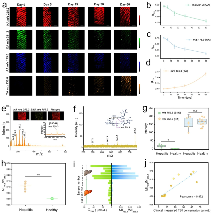
On the opposite aspect, we noticed that the age of the fingerprint was one other evident piece of data to slender down the individuals of curiosity. Stimulated by the great efficiency proven above, an try was undertaken to evaluate the age of the fingerprint utilizing the IVONSs:Sm-assisted LDI-MS instrument. For the sake of accuracy, all of the fingerprint samples have been saved at 30 °C and 60% relative humidity (RH). Determine 4g offered MS spectra of the consultant recent and aged fingerprints within the negative-ion mode. The unsaturated OA was discovered to endure peroxidization response, leading to a sign lower at m/z 281.2, whereas the MS alerts of saturated HA in fingerprints have been comparatively secure over time. On the identical time, two further peaks associated to the degradation of OA appeared with extended fingerprint age, which have been recognized as oleic acid hydroperoxide (OAHP, m/z 314.2) and 9-oxo-nonanoic acid (ONA, m/z 172.1). The peroxidization mechanism of OA based mostly on the previous research was given in Fig. 4h [43]. Specifically, OA undergoes a radical-mediated autoxidation in ambient air, forming isomerization merchandise of OAHP, secondary oxidation merchandise like ONA and nonanal are derived from the cleavage of OAHP. Likewise, the lower of the oxidizable AA and the brand new peak emerged at m/z 136.0, which was assigned to threonic acid (TA), confirmed the oxidative degradation pathway of AA. The broadly accepted mechanism of AA oxidation steered the technology of a collection of energetic intermediates (e.g. dehydroascorbic acid (DHA), diketogulonic acid (DKG)) and threonic acid (TA) [42, 44]. That’s, DHA, an oxidized type of AA, is then hydrolyzed to DKG, the intermediate DKG is additional decomposed to TA after the rearrangement response (Fig. 4i). The corresponding MS photographs have been compelling assist for these tendencies of OA and AA over time. The MS picture of HA spatial sample remained clearly seen and virtually invariable on the fingerprint ridges over time (Fig. 5a). In distinction, the fingerprint patterns steadily light for OA and AA from day 0 to day 60. The MS picture of TA was absent in the course of the preliminary interval of fingerprint growing old however turned a lot clearer with time happening. These outcomes gave a visible demonstration of the time-dependent modifications of the consultant LMW compounds in fingerprints. Importantly, quantitative evaluation of the modifications within the LMW compounds implied the potential for monitoring the age of fingerprints. To cut back systematic error, the MS alerts have been normalized by calculating the depth ratios (ROA, RAA, and RTA) utilizing the next equations: ROA = IOA/IHA, RAA = IAA/IHA, RTA = ITA/IHA, the place IOA, IAA, and ITA represented the MS sign intensities of OA at m/z 255.2, AA at m/z 175.1, and TA at m/z 136.0, respectively, and IHA stood for the HA depth at m/z 281.2 with a non-significant change. Therefore, Fig. 5bd illustrate the curves of ROA, RAA, and RTA over a two-month interval of fingerprint growing old, respectively. The lower of ROA (RAA) and the rise of RTA have been in keeping with the variations within the MS photographs in Fig. 5a. Equally, Determine S22b ~ d offered a constant pattern of the calculated MS depth ratios (ROA, RAA, and RTA) over time in comparison with Fig. 5b ~ d. The insignificant distinction amongst all of the teams steered a tiny affect of ambient fluctuations on the curves of ROA, RAA, and RTA, confirming the validity of the means of building fingerprint age. Furthermore, for a simulation experiment for fingerprint age dedication, ten simulated fingerprint specimens with totally different growing old occasions have been analyzed by way of the IVONSs:Sm-assisted LDI-MS strategy. Semi-quantitative analysis of fingerprint age was carried out based mostly on the time-dependent MS depth ratios (ROA, RAA, and RTA). Inference outcomes for fingerprint growing old time have been derived by adopting the typical growing old time of the three analytes. Comparisons between the true and predicted outcomes have been visualized by a warmth map, and the colour of the squares denoted the gradation of fingerprint age (Determine S23a). Verification with low prediction error proved the practicality of the IVONSs:Sm-assisted LDI-MS instrument for figuring out the growing old time of unknown fingerprint samples.
(a) IVONSs:Sm-assisted LDI-MS photographs of recent and aged fingerprints in negative-ion mode, and time-dependent MS depth ratios of (b) OA, (c) AA, and (d) TA. (e) Consultant IVONSs:Sm-assisted LDI-MS spectra of fingerprints from a hepatitis affected person. The inset reveals the corresponding spatial distribution of HA and BilG. (f) The MALDI LIFT-TOF/TOF MS/MS spectrum of BilG obtained from a fingerprint pattern in negative-ion mode. (g) Field plot of MS sign depth at m/z 255.2 and 759.3 between wholesome volunteers and hepatitis sufferers (* P < 0.05). (h) Scattered dot plots of the calculated MS sign depth ratio (MI759.3/MI255.2) between wholesome volunteers and hepatitis sufferers (* P < 0.01). (i) Comparability between MI759.3/MI255.2 and clinically measured TBil focus from 6 hepatitis sufferers (No. 1 ~ 6) and 10 wholesome volunteers (No. 7 ~ 16). (j) Correlation between MI759.3/MI255.2 and clinically measured TBil focus from 6 hepatitis sufferers and 10 wholesome volunteers. Scale bars in (a) and (e) signify 1 mm
We subsequently sought to increase the applying of the IVONSs:Sm-assisted LDI-MS technique to observe biomarkers excreted from finger sweat glands. It was discovered that elevated ranges of free bilirubin (bilirubin glucuronide, BilG) within the biofluids is expounded to varied hepatopathies [45]. Therefore, quickly monitoring the extent of BilG in a label-free method is of nice significance for early prognosis (Determine S23b). With its dependable, extremely delicate, and label-free options, IVONSs:Sm-assisted LDI-MS is especially well-suited for hint BilG detection. The everyday MS spectrum of the sweat fingerprint pattern from acute hepatitis sufferers displayed the [M − H]− peak of BilG at m/z 759.3, which was putatively recognized by way of MALDI LIFT-TOF/TOF MS (Fig. 5e, f). Moreover, the MS photographs exhibited a sweat pore-centered spatial distribution of BilG within the fingerprint ridge space (inset, Fig. 5e). For the sweat fingerprint pattern from a wholesome donor, a MS spectrum and not using a BilG sign was noticed (Determine S23c). To additional consider the feasibility of the proposed IVONSs:Sm-assisted LDI-MS instrument in medical evaluation, BilG ranges in fingerprints from 10 wholesome donors and 6 hepatitis sufferers have been analyzed. Determine S24 enumerated the negative-ion mode MS spectra of fingerprints from the hepatitis affected person group, and better MS depth at m/z 759.3 than the wholesome group identified the rise of BilG focus within the sweat of hepatitis sufferers (Fig. 5g and Determine S25). Through the use of the MS sign of HA (m/z 255.2) as an inside commonplace, this discrepancy may very well be extra clearly discriminated by way of a ratiometric parameter (MI759.3/MI255.2), the place MI759.3 and MI255.2 represented the MS sign depth at m/z 759.3 and 255.2, respectively (Fig. 5h). Moreover, the serum whole bilirubin (TBil) degree of the 16 members, which was broadly utilized in serodiagnosis, was decided by the clinically bilirubin oxidase technique. Determine 5i confirmed a superb consistency between the parameter MI759.3/MI255.2 and TBil focus in figuring out hepatitis sufferers, which was additional corroborated by Pearson correlation evaluation (r = 0.872) in Fig. 5j. As a result of superiorities of the label-free method, non-invasive sampling, and fast response, the proposed technique possesses excellent benefits over many bilirubin assay methods [46, 47].


