Synthesis and formation mechanism of functionalized HA hydrogels
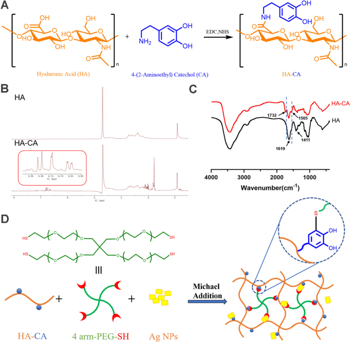
As proven in Fig. 1A, CA was grafted onto HA by EDC/NHS chemistry to supply the conjugate HA-CA. The copolymer construction was validated by 1H NMR spectra and FTIR spectrum, as proven in Fig. 1B and C. The height noticed at δ 1.8–2.0 corresponds to the proton in methylene of HA. The conjugation of CA to HA was confirmed by the presence of aromatic-proton peaks at δ 6.70–6.85 ppm and methylene-proton peaks at δ 3.1 and a pair of.8 ppm. In response to the calculation, the substitution diploma of CA in HA was 12.9%. Compared with the FTIR spectrum of HA, the absorption peak at 1732 cm−1 might assigned to the stretching vibration peak of − COOH. As well as, the uneven and symmetric stretching vibration intensities of − COONa at 1619 cm−1 and 1411 cm−1 have been considerably decreased. Much more vital, HA-CA confirmed a stronger absorption peak at 1565 cm−1, which was shaped by N–H bending vibration of amide II band. These findings affirm profitable conjugation between HA and CA by way of amide bonds. The schematic of HP-Ag construction is proven in Fig. 1D. CA loses two electrons and two protons to turn into quinone, which reacts extremely with the mercaptan group by means of the Michael addition response. CA teams can type bodily or chemical bonds with completely different surfaces, and quinone teams promote the cohesion of hydrogels [28].
Physicochemical properties of the hydrogel
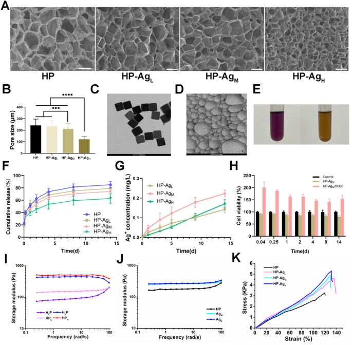
The micromorphology and common pore dimension of freeze-dried hydrogels with completely different Ag NPs contents are proven in Fig. 2A and B. All hydrogels exhibit a uniform community construction and porous interpenetration. The crosslinking density might be mirrored by hydrogel pore dimension to some extent. With the rise of Ag NPs content material, the pore dimension of hydrogel progressively decreases. The typical pore sizes of HP, HP-AgL, HP-AgM and HP-AgH have been 242 ± 54.4 μm, 234 ± 48.3 μm, 211 ± 46.2 μm and 122 ± 25.3 μm, respectively. The porous construction permits the hydrogel to soak up blood and exudate from the wound tissue, offering sufficient house for cell proliferation and thus dashing up hemostasis [29]. The addition of Ag NPs enhances the gel community construction and occupies the ordered pores within the hydrogel, resulting in a discount in pore dimension. Two doable causes of gel community enhancement are: First, CA on the HA-CA polymer deprotonates to type quinone teams, which assist the deposition of HA-CA on the Ag floor by means of hydrophobic motion; Secondly, sulfophilic Ag can react with obtainable sulfhydryl teams in 4-arm PEG-SH to type Ag–S bond, which connect the PEG-SH to Ag NPs floor [30,31,32].
Physicochemical properties of HP-Ag/bFGF hydrogel. A Scanning electron microscope photos of the freeze-dried HP-Ag hydrogel. Scale bar: 500 μm. B The pore dimension of the hydrogels ready by completely different concentrations of Ag NPs. C Morphology of of Ag NPs. D TEM micrograph of Ag NPs in HP-Ag hydrogel. E DPPH radical scavenging charge of HP hydrogel. F Cumulative launch curve of bFGF in HP-Ag/bFGF hydrogel. G Cumulative launch curve of Ag+ in HP-Ag hydrogel. (H) Bioactivity of launched bFGF. I G′ of HP hydrogels with completely different matrix contents measured in frequency sweep. J G′ of HP-Ag hydrogel measured in frequency sweep. Ok Compressive stress–pressure curves of HP-Ag hydrogel. Knowledge are offered as imply ± S.D, n = 3
Ag NPs have a big particular floor space, and their antibacterial exercise is affected by their particle dimension. From Fig. 2C and Further file 1: Fig. S1, it may be noticed that Ag NPs are in common dice form with a particle dimension of 100 nm and a possible of − 23.87.
As proven in Fig. 2E, the DPPH-clearance of the HP hydrogel was 59.53%. The clearance was decrease than anticipated, contemplating that there have been fewer CA teams obtainable on HA, as a few of them might have been oxidized to quinone or connected to different teams.
The discharge habits of bFGF in HP-Ag/bFGF was studied, and it may be clearly noticed from Fig. 2F that bFGF was launched quickly in HP hydrogel, whereas the incorporation of Ag NPs slowed down the discharge charge. For HP hydrogel, a burst launch of bFGF (51.3%) was noticed on day 1, whereas 46.2% and 39.9% bFGF launch have been noticed in HP-AgM and HP-AgH hydrogels, respectively. The discharge in HP hydrogel reached 73.9% on day 4, and the discharge charge in HP-AgM and HP-AgH hydrogels have been 64.4% and 53.4%, respectively. These items of proof counsel that long-term secure launch might be achieved by including Ag NPs to the hydrogel to boost the community construction, thus simplifying drug administration.
The antibacterial exercise of HP-Ag hydrogel relies on the discharge of Ag from the hydrogel into the contaminated website. Ag NPs can’t solely cross-link networks in hydrogel, but in addition act as reservoirs for Ag. As proven in Fig. 2G, a gradual and sustained launch of Ag was noticed all through the take a look at time. The quantitative methodology of Ag launch used doesn’t permit the differentiation of its type (ion or nanoparticle) [33]. The cumulative Ag releases of the HP-AgL, HP-AgM and HP-AgH after 14 days have been 0.143 mg L−1, 0.203 mg L−1 and 0.171 mg L−1, respectively. The comparatively excessive launch on the primary day ensured a powerful antibacterial impact in the course of the preliminary section of tissue therapeutic, and a specific amount of Ag remained on the wound website though the discharge of Ag was decreased later [34]. The present launch was barely decrease than anticipated [35,36,37], presumably as a result of some Ag NPs participated within the community as a crosslinking agent, and the floor of Ag NPs was connected to HA-CA or 4-arm PEG-SH, thus delaying Ag launch. This low focus ensures efficient sterilization above subinhibitory concentrations to keep away from the formation of bacterial biofilms, whereas being protected for cells and tissues [32, 33, 36].
Typically, Ag NPs are likely to accumulate and might result in giant will increase in native Ag concentrations, which can have an effect on cell exercise. The consequence reveals that Ag NPs have been distributed uniformly in HP-Ag (Fig. 2D). It’s speculated that the hydroxyl and sulfhydryl teams deposited on the floor of Ag NPs trigger the secure and uniform distribution of Ag NPs within the community construction.
The bioactivity of launched bFGF was evaluated by the stimulating impact on NIH/3T3 cell viability. From Fig. 2H, in contrast with upkeep medium group and HP-Ag group, the cell viability of the bFGF launch group was considerably elevated, indicating that bFGF in hydrogel maintained bioactive.
The dynamic rheological evaluation was performed to research the mechanical efficiency of hydrogels. Determine 2I and J reveals the storage modulus (G′) of hydrogels with completely different contents of HA-CA, 4-arm PEG-SH and Ag NPs measured in frequency sweep. All hydrogels confirmed elastic solid-like hydrogel after gelation. The G’ of hydrogel elevated with the rising focus of HA-CA and 4-arm PEG-SH. As a result of introduction of HA-CA and 4-arm PEG-SH, the community cross-linking of hydrogels was enhanced by means of a number of interactions, and the stiffness of hydrogels was improved. Equally, the G’ of HP-Ag hydrogel progressively elevated with the rise of Ag NPs content material. The G’ elevated with the rise of pressure frequency throughout the angular frequency of 0.1–100 rad s−1. The rise in modulus with pressure frequency is the strain-stiffening habits of hydrogels[38]. This property allows hydrogel to supply a greater bonding impact when excessive pressure and robust stress are generated on the bonding website[39].
Compressive stress–pressure assessments have been carried out on the hydrogels to evaluate its mechanical power (Fig. 2Ok). According to the rheological outcomes, the stresses of HP-Ag/bFGF hydrogels have been larger than that of HP hydrogel. Particularly, the compressive power of HP hydrogel was as little as 3.3 kPa, whereas the compressive power of HP-AgL, HP-AgM and HP-AgH elevated by 4.7 kPa, 4.9 kPa and 5.3 kPa, respectively. With the rise content material of Ag NPs, the compressive rupture pressure of hydrogel additionally elevated barely. The compressive rupture pressure of HP hydrogel was 119.7%, and that of HP-AgL, HP-AgM and HP-AgH have been 133.6%, 129.3% and 130.9%, respectively. From Fig.S2A, the HP hydrogel has a further weight reduction temperature vary in comparison with the load lack of HA, with full thermal decomposition occurring on the 300–420 °C stage. From Fig.S2B, the entire residual weight of HP-AgH was at all times larger than that of the HP-AgL composite system at every weight reduction stage. These outcomes point out that Ag NPs are embedded within the hydrogel matrix and the hydrogels with larger Ag NPs content material can type extra secure constructions. In abstract, these outcomes point out that HP-Ag/bFGF hydrogel holds a number of deserves together with porous microstructure, bFGF launch regulation, Ag launch and adjustable stiffness functionality.
in vitro biocompatibility and antibacterial exercise
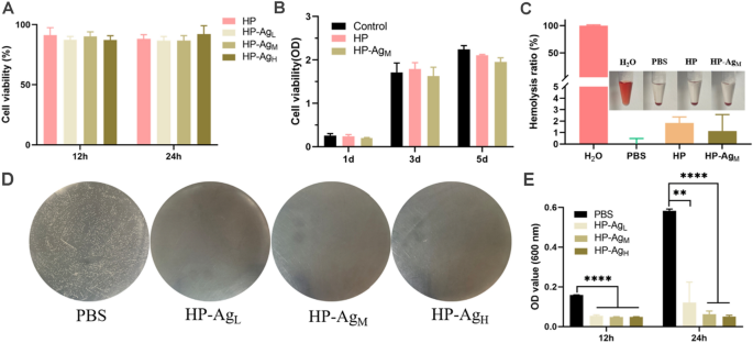
The flexibility of hydrogels impressed us to discover the organic properties required for his or her software in wound dressings. Good biocompatibility is an important prerequisite for hydrogel dressings as a result of they’re in direct contact with tissues and blood in sensible purposes. The cytotoxicity of HP-Ag hydrogel on NIH/3T3 was evaluated utilizing a CCK-8 assay in vitro, with PBS because the management group. As proven in Fig. 3A, after 12 h and 24 h tradition, the relative cell development charge of every group of hydrogels exceeded 86%, far larger than the minimal non-toxic commonplace of 70%. Comparable outcomes have been obtained in proliferation experiments (Fig. 3B). There was no important distinction in cell proliferation charge between the HP-Ag hydrogel and management group, indicating that hydrogels had good cytocompatibility. After 3 days and 5 days of incubation, the cell exercise of HP-AgM decreased barely in contrast with PBS group, which can be as a result of inactivation of some cells uncovered to residual Ag NPs. Most typical cross-linkers utilized in catechol polymers, similar to Fe3+ and NaIO4, are cytotoxic, limiting their use as biomaterials. HP-Ag ready utilizing 4-arm PEG-SH and Ag NPs as cross-linkers had no cytotoxicity and is predicted for use in scientific wound dressings.
Biocompatibility and antibacterial properties of hydrogels. Cell viability of NIH/3T3 co-cultured with hydrogel extracts for A 12 h and 24 h. and B 1d, 3d and 5d. C Hemolytic exercise assay of HP and HP-AgM hydrogel. Inset is the corresponding {photograph}. D Pictures of survival micro organism colonies (S. aureus, white dots in plates) rising on agar plates after direct contact incubation with HP and HP-Ag hydrogels. PBS was used as management. E OD values at 600 nm of S. aureus answer co-cultured with the hydrogels for 12 h and 24 h. All information are offered as imply ± SD, n = 4, **p < 0.01, ***p < 0.001, ****p < 0.0001
Hemocompatibility is a vital issue to guage the compatibility between blood and biomaterials. The usual hemolysis exercise take a look at was used to guage the hemolysis ratio between homogeneous hydrogel and pink blood cells (RBCs). As proven in Fig. 3C, hemolysis charges of HP and HP-AgM hydrogels have been 1.83% and 1.13%, respectively. The hemolysis charges have been just like that of the PBS damaging management, however a lot decrease than that of the optimistic management and nicely under the allowable restrict of 5%. Direct visible commentary confirmed that the supernatant of the hydrogel group was yellowish and nearly indistinguishable from the PBS group. Within the optimistic group, the supernatant was vivid pink brought on by RBCs lysis, indicating that the hemolysis of HP and HP-AgM hydrogels was negligible. The low hemolysis charge is as a result of hydrophilicity of HA and 4-arm PEG-SH, which reduces RBCs destruction.
The antibacterial exercise of HP-Ag hydrogel towards gram-positive S. aureus, the consultant reason for pores and skin infections, was evaluated by colony counting. As proven in Fig. 3D, numerous bacterial colonies appeared within the agar plate of PBS group, whereas no viable colonies appeared in HP-Ag teams, indicating sturdy bactericidal results. Equally, OD values of the S. aureus options co-cultured with HP-Ag hydrogel have been considerably decrease than that in PBS group, and the suspensions have been clear after 12 and 24 h (Fig. 3E). These outcomes counsel that HP-Ag hydrogel was efficient in killing S. aureus and had negligible harm to cells. It needs to be emphasised that the extremely efficient antibacterial properties of HP-Ag hydrogel additionally drastically scale back the unintended effects of wound therapy which may be brought on by excessive doses[40, 41]. As for doable antibacterial mechanisms, it’s speculated that first the obtainable phenol hydroxyl teams in HP-Ag hydrogel entice the micro organism, after which the positively charged Ag+ from Ag NPs work together electrostatic with the phosphoric teams of the phospholipids within the cell membrane, killing the micro organism by interfering with DNA replication and denaturing microbial proteins [42,43,44]. General, HP-Ag hydrogel confirmed superior cytocompatibility, hemocompatibility and robust antibacterial exercise, which has nice potential for scientific software.
Speed up wound therapeutic means of acute wound
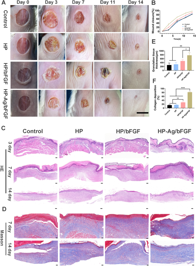
The wound therapeutic processes of acute full-thickness pores and skin defects have been monitored photographically (Fig. 4A), and the corresponding calculation of the present wound space have been offered in Fig. 4B. The wound dimension of every group confirmed a lowering development over time. After 14 days of therapy, the wound space of HP-Ag/bFGF therapy was the smallest, and the wound turned easy and a few new epidermal and dermal tissues showing. In distinction, the pores and skin floor of the wound in different teams was pink with apparent scabs, suggesting the formation of early scar tissues. The redness of those scars is often because of incomplete angiogenesis [19]. Wound closure diverse considerably in the course of the first 7 days after modeling because of self-contraction of the pores and skin. The wound closure space of the management group (6.6%) was considerably decrease than that of the HP-Ag/bFGF group (37.7%) on day 3 and was solely 48.7% on day 7. The wound closure charge of HP-Ag /bFGF group reached 68.2%, larger than that of HP/bFGF group (54.7%). In contrast with different teams, the HP-Ag/bFGF composite hydrogel group was more practical in selling wound therapeutic. On the one hand, the hydrogel shaped in situ can match the wound nicely and cling carefully to the wound website to keep away from microbial an infection. However, the water-retaining potential of HA preserves the moist surroundings required for wound therapeutic, and HP-Ag/bFGF can sustainably present bFGF for tissue regeneration.
A Pictures of the pores and skin wounds of varied teams on day 0, 3, 7, 11and 14 (Scale: 10 mm). B Wound closure evaluation from the present wound space on day 3, 7, 11 and 14. C H&E staining of the wound sections on day 3, 7 and 14 (Scale: 100 μm). D Masson staining of the wound sections on day 7 and 14 (Scale: 50 μm). E Semi-quantification of pores and skin granulation tissue thickness on day 7. F Collagen quantity fraction of the wound sections on day 7. All information are offered as imply ± SD, *p < 0.05, **p < 0.01, ****p < 0.00001, n = 3
With a view to observe the method of wound therapeutic extra straight and precisely, the histological modifications of pores and skin have been evaluated. H&E staining photos (Fig. 4C) present that there was no or solely a skinny layer of granulation tissue underneath the pores and skin of the management, HP and HP/bFGF teams, and numerous inflammatory cells infiltrated. The semi-quantitative evaluation (Fig. 4E) reveals that the granulation tissue thickness in HP-Ag/bFGF group was 776 μm, which was clearly larger than that in different teams. On day 14, the brand new dermis was clearly thickened and uneven in management group, and the inflammatory cells have been clearly gathered underneath the dermis, each of which might result in the formation of scar on the wound website [45, 46]. Nonetheless, the HP-Ag/bFGF group confirmed uniform epidermal tissue thickness, orderly granulation tissue, no evident irritation occurred, and the regenerated dermis tissue with appendages like hair follicles and sebaceous glands was detected, all of which have been vital indicators of pores and skin scarless therapeutic.
Because the help of cell development, collagen can promote the proliferation and differentiation of tissue cells and create a greater microenvironment. Nonetheless, extreme manufacturing and disorderly deposition of collagen within the dermis can result in scar manufacturing [5, 47]. Masson staining was used to guage collagen deposition at wound websites (Fig. 4D). The collagen deposition quantity of HP-Ag/bFGF group was larger than that of different teams on day 7, and the collagen fibers have been loosely and well-organized distributed in dermal tissue on day 14. That is according to the traits of scarless pores and skin restore, the place collagen accumulates in giant portions within the early phases after which partially fades and rearranges into the tissue [48]. Semi-quantitative evaluation (Fig. 4F) reveals that the collagen deposition was solely 15.2% in management group on day 7, whereas it was 81.7% in HP-Ag/bFGF group, considerably larger than that in different teams. These outcomes point out that HP-Ag/bFGF composite hydrogels can speed up wound regeneration and promote wound scarless therapeutic.
TNF-α, a typical proinflammatory cytokine, was chosen to guage the anti-inflammatory impact of hydrogels in vivo. The expression of TNF-α on the wound website on day 3 was proven in Further file 1: Fig.S3 and Fig.S4. The expression of pro-inflammatory components in management group was considerably larger than that in HP/bFGF and HP-Ag/bFGF group. As well as, TNF-α expression was lowest in HP-Ag/bFGF group, which was associated to the continual launch of Ag to supply an anti-inflammatory micoenvironment. As proven in Further file 1: Fig.S5, HP /bFGF and HP-Ag/bFGF group confirmed extra mature blood vessels within the wound mattress than the opposite teams. Lengthy-term launch of bFGF and Ag promotes the formation of mature blood vessels and the regression of immature blood vessels, thus offering sufficient oxygen, vitamins, and development components for tissue regeneration.
Reworking development issue TGF-β can induce fibroblasts to distinguish into myofibroblasts, which is carefully associated to scar formation in tissues. From Further file 1: Fig.S6 and S7, the expression of TGF-β in HP-Ag/bFGF group was considerably decrease than that in different teams. These outcomes point out that HP-Ag/bFGF composite hydrogel can facilitate scarless wound therapeutic by down-regulating the expression of TGF-β.
Analysis of bacteria-infected wound therapeutic in vivo
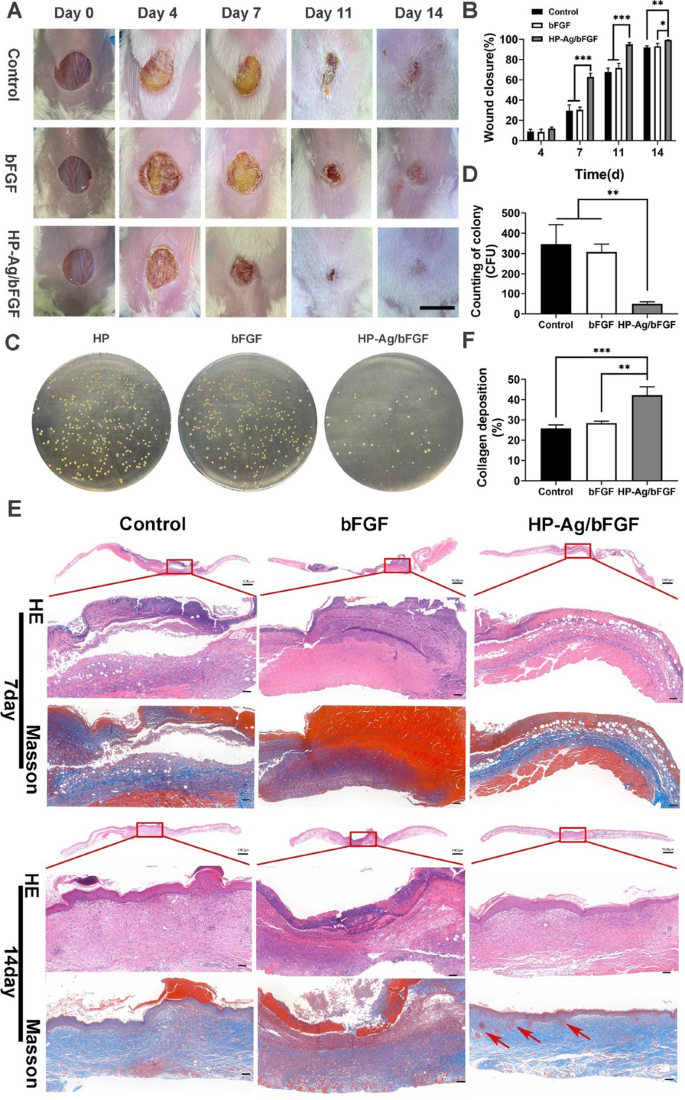
The HP-Ag/bFGF hydrogel has the flexibility to constantly launch Ag, and in vitro experiments have proven that it has a powerful antibacterial impact. To additional increase its scientific software, a bacterial contaminated wound mannequin was used. Determine 5A reveals that S. aureus on the wound website of the management group and the bFGF group grew wantonly. From day 4 to day 7, the wound was coated by numerous bacterial colonies, which critically hindered its closure course of. There was no important change within the charge of wound closure till the bacterial layer and blood scab have been shed. Nonetheless, the HP-Ag/bFGF group skilled a extra common closure course of and was much less affected by bacterial an infection. On day 14, the wound was easy and closed and even vanished for therapy group of HP-Ag/bFGF, whereas the wound boundaries have been nonetheless noticed for different teams. Within the means of pores and skin restore, the wound closure charge of HP-Ag/bFGF group was considerably larger than that of different teams (Fig. 5B).
A Pictures of the pores and skin wounds of varied teams on day 0, 4, 7, 11and 14 (Scale: 10 mm). B Wound closure evaluation from the present wound space on day 4, 7, 11 and 14. C Pictures of survival micro organism colonies (S. aureus, yellow dots in plates) rising on agar plates from wound websites on day 7 after completely different remedies. D Counting of survival micro organism colonies rising on agar plates from wound websites on day 7. E H&E and Masson staining of the wound sections on day 7 and 14. (Scale: 100 μm). F Collagen quantity fraction of the wound sections on day 7 (pink arrow: hair follicles). All information are offered as imply ± SD, **p < 0.01, ***p < 0.001, n = 3
On day 7, pores and skin tissues have been homogenized and diluted at an applicable ratio earlier than being smeared on the agar plate. Pictures and colony counts of contaminated pores and skin in numerous teams are proven in Fig. 5C and D. The variety of colonies within the HP-Ag/bFGF group was considerably lower than that within the different two teams, which was according to the outcomes of in vitro antibacterial experiments. These outcomes affirm that HP-Ag/bFGF hydrogel was able to destroying bacterial as an antibacterial platform, which is according to the outcomes of earlier research [49].
As proven in Fig. 5E, just like the acute wound, the dermis in management group was thickened, with mastoid processes, and fibroblasts have been considerable and irregularized. In the meantime, the collagen fibers have been quite a few, dense and thick, with disordered association, and the boundary between dermal reticular layer and epidermal layer was blurred. In distinction, the epidermal layer in HP-Ag/bFGF group was flat and skinny, and there are fewer fibroblasts, and most of them are organized in parallel. A key characteristic of optimum wound therapeutic is reworking the ECM by depositating collagen in a well-organized community to revive the traditional construction of tissue. The unfastened and orderly association of collagen and the presence of regenerated pores and skin appendages similar to hair follicles and sebaceous glands additional affirm that HP-Ag/bFGF results in scarless and efficient regeneration. Collagen content material within the HP-Ag/bFGF group was considerably larger than that within the different two teams on day 7, as proven in Fig. 5F.
Regeneration and reworking of the blood provide system is vital for tissue regeneration. New blood vessels can present needed vitamins and oxygen provide for tissue reconstruction and carry away metabolic waste in time [50]. CD31 and VEGF have been detected to guage wound angiogenesis. As proven by the CD 31 staining leads to Fig. 6A, the immature capillaries within the management group have been dense and disordered. Determine 6B and C present that HP-Ag/bFGF comprised considerably extra mature vessels than the opposite two teams, whereas the briefly constructed immature vessels degenerated over time. This management of vessel density by partially blocking capillary development might result in a decreased however absolutely practical vascular system, which has been proven to enhance long-term therapeutic outcomes and keep away from scarring [51, 52].
A Immunohistochemistry staining photos of CD31(pink arrow: mature blood vessels) and VEGF within the wound tissues on day 14. Semi-quantitative evaluation of the expression degree of CD31-positive ( +) mature vessels B, VEGF C within the wound tissues on day 14. D Immunohistochemistry staining photos of TGF-β and α-SMA within the wound tissues on day 14. Semi-quantitative evaluation of the expression degree of TGF-β E and α-SMA F within the wound tissues on day 14 (Scale: 100 μm). All information are offered as imply ± SD. *p < 0.05, **p < 0.01, n = 3
TGF-β straight induces α-SMA expression in recruited fibroblasts, selling myoblast differentiation into myoblasts, which in flip promotes uncontrolled ECM manufacturing, resulting in scarring [53]. Subsequently, TGF-β exercise was related to elevated scar formation and fibrosis induction on the later stage of wound therapeutic. Determine 6D and E present that the expression of TGF-β in HP-Ag/bFGF group was considerably lower than that of the opposite two teams. These outcomes counsel that HP-Ag/bFGF has better potential to advertise scarless wound therapeutic. The low expression of α-SMA in Fig. 6F additionally confirmed this conclusion. As well as, as proven in Further file 1: Fig.S8 and Fig.S9, the consultant organs (coronary heart, liver, spleen, lung, and kidney) have been examined by H&E staining, and no apparent pathological abnormalities or harm have been discovered within the organ sections, verifying the biosafety of HP-Ag/bFGF hydrogel for wound therapeutic.
Modulated macrophages polarization and irritation response
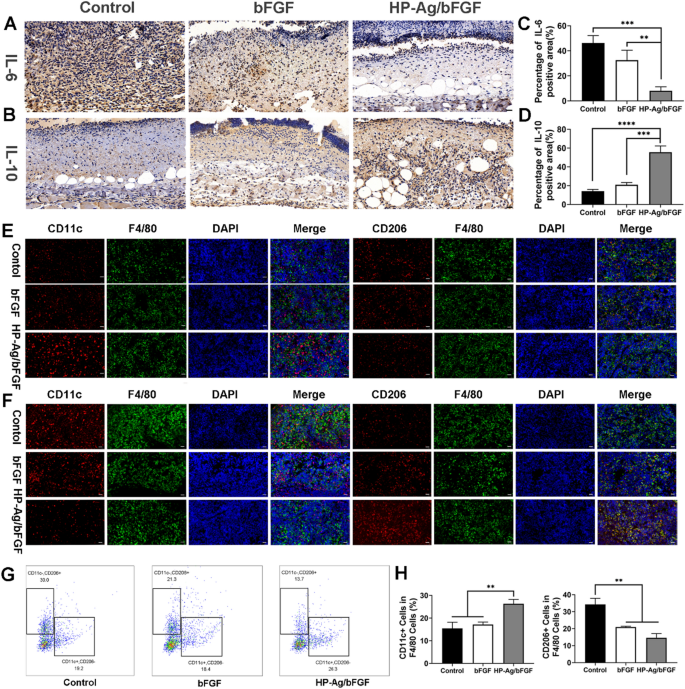
Interleukin (IL) is a bunch of cytokines that play a vital function within the inflammatory course of. IL-6, a pro-inflammatory cytokine, is launched early in irritation and has been proven to be an efficient stimulant of fibroblast proliferation [54]. IL-10, an anti-inflammatory and antifibrotic cytokine, has been reported to scale back the expression of IL-6, IL-8, and different inflammatory genes, and considerably enhance collagen patterns [55, 56]. The content material of IL-6 in tissues of HP-Ag/bFGF group was considerably decrease than that in different teams, as proven in Fig. 7A and C. In the meantime, the content material of IL-10 in tissues of HP-Ag/bFGF group was considerably larger than that in different teams (Fig. 7B and D). These outcomes counsel that in contrast with the opposite teams, the tissue irritation was the weakest in HP-Ag/bFGF group on day 4 and the anti-inflammatory impact was the strongest on day 7.
Regulation of irritation and immune microenvironment. A Immunohistochemistry staining photos of IL-6 within the wound tissues on day 4. B Immunohistochemistry staining photos of IL-10 within the wound tissues on day 7. Semi-quantitative evaluation of the expression degree of IL-6 C and IL-10 D. Immunofluorescence staining of M1 and M2 sort macrophage within the spleen of contaminated mice on day 4 E and day 7 F. Multicolor stream cytometry G and quantitative evaluation H to detect the M1-type and M2-type macrophages within the spleen of contaminated mice on day 4. Scale bar: 20 μm (n = 3, **p < 0.01, ***p < 0.001, ****p < 0.0001)
Innate immunity is an important element of the physique’s protection towards pathogen invasion and performs an important function in mediating tissue restore. The transformation of macrophages from M1 phenotype to M2 phenotype is believed to be needed for wound therapeutic [57]. On day 4 of acute wound an infection, completely different macrophages subtypes have been labeled by immunofluorescence and multicolor stream cytometry. M1-type macrophages (F4/80 + /CD11c +) are pro-inflammatory immune cells that secrete inflammatory cytokines in the course of the acute section of an infection, whereas M2-type macrophages (F4/80 + /CD206 +) have anti-inflammatory and restore results [58]. From Fig. 7E, the variety of M1-type macrophages of HP-Ag/bFGF group was larger than that of the opposite two teams. Equally, stream cytometry confirmed the outcomes. As might be seen from Fig. 7G and H, M1-type macrophages within the HP-Ag/bFGF group have been considerably larger than these within the different two teams, whereas M2-type macrophages have been considerably decrease than these in management group. The outcomes point out that the immune response within the management group and the bFGF group was comparatively gradual on the preliminary stage of wound an infection, whereas the HP-Ag/bFGF group efficiently stimulated the immune response by means of the discharge of Ag, accelerating the method of irritation after an infection and harm. Determine 7F reveals the staining of M1-type and M2-type macrophages labeled by immunofluorescence 7 days after the an infection. Presently, with the progress of tissue restore, the variety of M2-type macrophages in HP-Ag/bFGF group was larger than that in different teams, whereas the variety of M1-type macrophages was the bottom. The outcomes point out that the inflammatory interval of the contaminated pores and skin tissue within the HP-Ag/bFGF group had transitioned to the following tissue restore interval, whereas the management group and the bFGF group have been nonetheless experiencing the inflammatory interval. This commentary is according to earlier immunohistochemical outcomes of IL-6 and IL-10.


