Synthesis and characterization of various Au NPs
The synthesis of Au NPs by a inexperienced one-pot methodology is a broadly accepted and continuously employed method recognized for its environmentally pleasant, uncomplicated, and cost-effective traits [37]. This methodology principally entails combining tetrachloroauric acid (HAuCl4) with a decreasing agent inside the similar container to provide natural or inorganic Au NPs [38]. The usual method for producing Au NPs employs sodium borohydride (NaBH4) because the decreasing agent [39]. Earlier investigations have highlighted that reducible purposeful teams like hydroxyl, amino, and amide teams in natural compounds can effectuate the discount of HAuCl4, yielding adorned Au NPs exhibiting different functionalities [40]. In our research, we noticed that NAS share related chemical buildings.
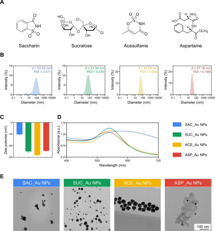
On this research, we synthesized SAC-decorated Au NPs (SAC_Au NPs), SUC-decorated Au NPs (SUC_Au NPs), ACE-decorated Au NPs (ACE_Au NPs), and ASP_Au NPs by combining SAC (0.05 mmol), SUC (0.05 mmol), ACE (0.05 mmol), ASP (0.05 mmol) (Fig. 1A), and HAuCl4 (0.05 mmol). Because of the distinctive reducibility of NAS, the Au3+ ions in HAuCl4 bear discount to Au0 by Au-O or Au–N bonds, facilitating the formation of the aforementioned adorned Au NPs. Residual NAS inside the system underwent elimination by dialysis. Dynamic mild scattering (DLS) measurements revealed that SAC_Au NPs, SUC_Au NPs, ACE_Au NPs, and ASP_Au NPs exhibited common sizes of 52.52, 51.34, 42.56, and 27.18 nm, respectively, with polydispersity indices (PDIs) of 0.511, 0.270, 0.258, and 0.199 (Fig. 1B). This implies the presence of small and uniformly dispersed sizes among the many 4 Au NPs. As depicted in Fig. 1C, SAC_Au NPs, SUC_Au NPs, ACE_Au NPs, and ASP_Au NPs displayed common zeta potentials of − 10.2, − 24.9, − 28, and − 24.4 mV, respectively, indicative of negatively charged nanoparticles with commendable stability. The UV–seen spectra exhibited absorption peaks inside the 500–600 nm vary, similar to the distinctive floor plasmon resonance of Au NPs (Fig. 1D). The UV–seen spectra of the Au NPs diminished by NaBH4 are proven inAdditional file 1: Determine S1.The noticeable redshift within the absorption peaks of the 4 NAS-decorated Au NPs (NAS_Au NPs) in comparison with these of the NaBH4-reduced Au NPs, which confirms the profitable ornament of NAS on the gold nanoparticles [41]. The scale, PDI, and zeta potential of NaBH4-reduced Au NPs are proven inAdditional file 1: Determine S2. Transmission electron microscopy (TEM) photos (Fig. 1E) unveiled uniform morphology and diminished particle sizes for SAC_Au NPs, SUC_Au NPs, ACE_Au NPs, and ASP_Au NPs, roughly measuring 30, 28, 35, and 12 nm, respectively. The TEM sizes proved smaller than the DLS sizes, as DLS displays hydrodynamic measurement whereas TEM evaluates the dimensions of dry particles [42]. Notably, smaller-sized Au NPs have demonstrated enhanced antimicrobial exercise [43], and ASP_Au NPs rising because the standout among the many 4 Au NPs because of their stability, well-defined round form, minimal measurement, and uniform dispersion. Additional, the extent of NAS certain to the AuNPs was measured utilizing the ortho-pthaldehyde (OPA) fluorescence assay [44]. As proven in Further file 1: Desk S1, it seems that roughly half of the NAS have certain to the Au NPs. OPA reacts with main amines to provide fluorescence, and the decrease binding effectivity noticed could also be because of some NAS collaborating within the discount course of, whereas others are concerned in conjugation. This might lead to alterations to the amino nitrogen teams, stopping their interplay with OPA. Moreover, we ready customary Au NPs using the NaBH4 discount methodology for subsequent experimental management.
Characterization of various Au NPs. A Molecular buildings of SAC, SUC, ACE, and ASP. B Particle measurement and dispersity of SAC_Au NPs, SUC_Au NPs, ACE_Au NPs, and ASP_Au NPs. C Zeta potential of SAC_Au NPs, SUC_Au NPs, ACE_Au NPs, and ASP_Au NPs. D UV–Vis spectra of SAC_Au NPs, SUC_Au NPs, ACE_Au NPs, and ASP_Au NPs. E TEM photos of SAC_Au NPs, SUC_Au NPs, ACE_Au NPs, and ASP_Au NPs
For in vitro antimicrobial exercise, antibiofilm results, biocompatibility, and in vivo mannequin research, the concentrations of SAC_Au NPs, SUC_Au NPs, ACE_Au NPs, and ASP_Au NPs had been stored in step with these of the Au NPs management group. Contemplating the challenges in figuring out post-dialysis concentrations of SAC, SUC, ACE, and ASP, their concentrations had been maintained at ranges much like these of the pre-dialysis SAC_Au NPs, SUC_Au NPs, ACE_Au NPs, and ASP_Au NPs teams. In all experiments, the Au NPs management group refers back to the group of NaBH4-reduced Au NPs.
Antimicrobial exercise of 4 NAS_Au NPs
As depicted in Further file 1: Desk S2, we chosen 30 distinctive scientific CRE strains and a couple of customary strains assessed their drug resistance profiles utilizing the microbroth dilution approach. Polymerase chain response (PCR) and sequencing methodologies had been employed to elucidate their resistance mechanisms, sure mechanism knowledge had been drawn from our prior research [45, 46]. The chosen strains exhibited resistance mechanisms spanning all 4 lessons of carbapenemases [47], encompassing Ambler class A (KPC, SHV, TEM, CTX-M, and so forth.), class B (NDM, IMP, and so forth.), class C (AmpC, CMY, and so forth.), and sophistication D (OXA). Further resistance mechanisms, akin to efflux pumps, OmpK37 mutations, and downregulation of OmpC and OmpF, had been additionally implicated. General, these strains displayed pronounced resistance to ertapenem (ETP) (≥ 64 µg/mL). Accordingly, we chosen ETP because the antibiotic management for subsequent experiments, sustaining its focus in step with that of the ASP_Au NPs group to underscore the scientific applicability of our formulated Au NPs in addressing CRE-related challenges.
As delineated in Desk 1, the MIC values of SAC_Au NPs, SUC_Au NPs, and ACE_Au NPs towards the 32 examined bacterial strains all exceeded ≥ 256 µg/mL, signifying an absence of notable antimicrobial exercise. Strikingly, ASP_Au NPs demonstrated distinctive antimicrobial efficiency towards all strains, displaying MIC values starting from 8 to 16 µg/mL, aside from CG1330, which exhibited an MIC worth of 4 µg/mL. The outstanding antimicrobial efficacy of ASP_Au NPs vis-à-vis the opposite three kinds of Au NPs could also be attributed to their uniform particle form, diminutive measurement, and the presence of ester, methyl, and phenyl moieties, which confer elevated hydrophobicity. Enhanced hydrophobicity facilitates the internalization of particles into bacterial cells [48]. Contemplating ASP_Au NPs as a member of the NAS_Au NPs group with distinctive antimicrobial results, we designated them for additional characterization and subsequent exploration encompassing antimicrobial exercise, antimicrobial mechanisms, antibiofilm exercise, biocompatibility, anti-inflammatory properties, and in vivo antimicrobial assessments.
Additional characterization of ASP_Au NPs
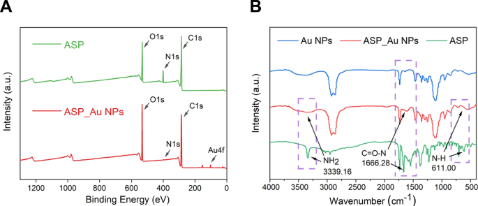
Given the potential of ASP_Au NPs in addressing scientific CRE challenges, we performed supplementary characterization by X-ray photoelectron spectroscopy (XPS) and Fourier-transform infrared spectroscopy (FTIR) to achieve deeper insights into ASP_Au NPs. As demonstrated within the XPS survey spectrum (Fig. 2A), ASP exhibited three discernible absorption peaks at roughly 530 eV, 400 eV, and 288 eV, correlating with the O1s, N1s, and C1s spectra, respectively. In distinction, ASP_Au NPs manifested three distinguished absorption peaks at roughly 530 eV, 288 eV, and 87 eV, similar to the O, C, and Au parts, respectively. A comparability of the 2 spectra unveiled the disappearance of the N component peak and the amplification of the Au component peak in ASP_Au NPs, suggesting the believable interplay between ASP’s amino and amide teams with Au, resulting in the institution of Au–N bond connections and consequently inflicting the obliteration of the absorption peaks that had been initially attributed to the amino and amide teams in ASP. To substantiate this speculation, we subsequently executed becoming of the Au 4f, N1s, and O1s spectra to scrutinize particular electron transfers through the chemical response (Further file 1: Determine S3). The Au 4f spectra (Further file 1: Determine S3A, B) exhibited two distinct absorption peaks for each ASP_Au NPs and customary Au NPs diminished by NaBH4, denoting Au 4f5/2 and Au 4f7/2. In view of the database and binding vitality positions, these peaks indicated the presence of Au0 formation. Moreover, ASP_Au NPs displayed a feeble absorption peak at round 85.78 eV, suggesting marginal Au1+ formation, presumably attributable to electron switch with -OH or nitrogen-containing teams. The N1s spectra (Further file 1: Determine S3C, D) displayed two distinguished absorption peaks for ASP situated at 400.14 eV and 401.57 eV, attributed to –NH2 and –NH–, respectively. In ASP_Au NPs, two absorption peaks emerged at 397.06 eV and 399.78 eV, additionally similar to –NH2 and –NH–, implying a shift within the nitrogen-containing group’s peak sample after loading Au onto ASP, signifying electron switch between Au and N, resulting in the institution of an Au–N bond. The O1s spectra (Further file 1: Determine S3E, F) unveiled conspicuous absorption peaks for ASP at 531.12 eV, 532.12 eV, and 533.88 eV, aligning with C = O, O = C–O, and C–OH, respectively. As compared, ASP_Au NPs exhibited absorption peaks at 531.88 eV, 532.48 eV, and 532.92 eV, ascribed to C = O, O = C–O, and C–OH, respectively. The one notable shift was noticed within the C–OH peak, suggesting electron switch between C–OH and Au after Au loading, possible because of the emergence of a small amount of Au1+.
The findings from FTIR analyses (Fig. 2B) indicated that the height patterns of ASP_Au NPs resembled these of Au NPs. ASP displayed a number of absorption peaks, with a definite peak at 1666.28 nm being the attribute peak of ASP [49]. Following Au loading, ASP_Au NPs exhibited conspicuous shifts in absorption peaks at wavelengths of 611 nm, 1666.28 nm, and 3339.16 nm, attributed to N–H, C = O–N, and –NH2 [50]. The disappearance of those three peaks implied the response between ASP’s amide construction (C = O–N–H) and amino group (–NH2) with Au, whereby the hydrogen on the nitrogen atom underwent substitution by Au to determine an Au–N bond, in step with the XPS evaluation. The distinct attribute peaks of Au NPs, ASP_Au NPs, and ASP are introduced in Further file 1: Determine S4. FTIR of the remaining three nano-cargos can be found within the Further file 1: Determine S5.
In vitro antimicrobial exercise research of ASP_Au NPs
To analyze the antimicrobial exercise of the uncooked supplies, we performed a microbroth dilution assay utilizing the 32 bacterial strains. We evaluated the antimicrobial results of ASP, Au NPs, and ASP_Au NPs individually. The outcomes, as proven in Desk 2, revealed that ASP and Au NPs alone had MIC values ≥ 256 µg/mL, indicating minimal antimicrobial exercise as anticipated. Nevertheless, ASP_Au NPs demonstrated outstanding antimicrobial efficiency. Though experiences recommend that NAS reveals some antimicrobial exercise at concentrations of 1500 µg/mL and above [51], we consider that this focus is far higher than the clinically used antibiotic doses and would possibly trigger sure negative effects [52], making them unsuitable to be used as antimicrobial brokers.
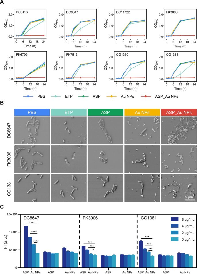
For additional insights into the consequences of ASP_Au NPs on CRE pressure development kinetics, chosen strains underwent development curve evaluation. As depicted in Fig. 3A, strains handled with phosphate-buffered saline (PBS), ETP, ASP, and Au NPs displayed regular development, whereas ASP_Au NPs therapy led to efficient antimicrobial motion, stopping bacterial development even after 24 h. The concentrations for every group had been beforehand specified. Moreover, to straight observe the morphological adjustments of micro organism after totally different therapies, a scanning electron microscopy (SEM) evaluation was carried out, and DC8647, FK3006, and CG1381 had been randomly chosen because the experimental strains. The SEM photos (Fig. 3B) revealed that micro organism handled with ASP_Au NPs on the MIC focus exhibited flattened and wrinkled morphology, together with distorted and ruptured cell our bodies. In distinction, the micro organism within the different therapy teams maintained common and intact mobile buildings. Elevated ROS ranges and membrane permeability adjustments are potential antimicrobial mechanisms of nanomaterials [53]. This aligns with our speculation, prompting us to conduct associated experiments. The ROS detection assay demonstrated that ASP_Au NPs considerably raised ROS ranges in strains DC8647, FK3006, and CG1381. The ROS enhance displayed a dose-dependent sample and even induced substantial ROS elevation in micro organism under the MIC focus (Fig. 3C). To substantiate that ROS is the core antimicrobial mechanism of ASP_Au NPs, we additional carried out ROS clearance experiments to research whether or not ASP_Au NPs nonetheless have vital antimicrobial exercise after clearing ROS. On this research, quercetin, a plant-derived pure flavonoid recognized for its highly effective antioxidant exercise and ROS scavenging properties [54, 55], was used because the ROS scavenger. After quenching ROS, the MIC values of ASP_Au NPs for a similar bacterial strains elevated from 4–16 µg/mL to ≥ 256 µg/mL, an MIC escalation of at the least 16–64 fold (Further file 1: Desk S3). This confirmed that elevated ROS ranges are the first antimicrobial mechanism of ASP_Au NPs. Moreover, we investigated the impression on inside membrane permeability. Within the propidium iodide (PI) membrane permeability assay (Further file 1: Determine S6), ASP_Au NPs at varied concentrations considerably elevated membrane permeability in comparison with the PBS management group (represented as 0 µg/mL), as evidenced by the corresponding rise in fluorescence depth. Moreover, ASP_Au NPs on the MIC focus (8 µg/mL) exhibited a pointy enhance in fluorescence depth, albeit to a lesser extent at sub-inhibitory concentrations, however nonetheless with vital statistical variations. This implies that ASP_Au NPs can intrude with bacterial morphology and regular physiological capabilities. Conversely, Au NPs and ASP at totally different concentrations didn’t exhibit a notable enhance in fluorescence depth. This discovering aligns with the noticed adjustments in bacterial morphology beneath scanning electron microscopy. Subsequently, membrane permeability may doubtlessly function a secondary antimicrobial mechanism of ASP_Au NPs.
The antimicrobial means of ASP_Au NPs in vitro and its core mechanism. A Development curves of scientific CRE strains after therapy with totally different formulations. B Consultant SEM photos of scientific CRE strains after therapy with totally different formulations. C ROS ranges in scientific CRE strains after therapy with totally different concentrations of formulations
Furthermore, to exclude potential synergistic results between ASP and Au NPs that may impression the research, a checkerboard assay was performed for validation. As depicted inAdditional file 1: Desk S4, inside the randomly chosen experimental strains, each the mixture therapy group and the monotherapy group exhibited MIC values of ≥ 256 µg/mL. This implies the absence of a big synergistic impact over a broad vary. This means that the antimicrobial efficiency of ASP_Au NPs originated from the “ornament” of ASP onto Au NPs, reasonably than a mere “synergistic” impact.
In vitro biofilm inhibition research of ASP_Au NPs
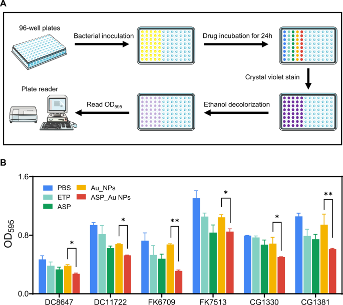
Bacterial biofilms are communities of micro organism connected to surfaces, offering inside micro organism with considerably higher antibiotic resistance, as much as 1000’s of occasions extra [56]. Within the context of hospital an infection management, biofilms shaped on medical gadgets and wound surfaces enormously impression affected person well being [57]. Moreover, inside the meals trade, microbial biofilms usually develop on meals packaging, posing dangers to meals security [58, 59]. Fortunately, quite a few research have reported the antimicrobial biofilm exercise of varied nanoparticles [60]. Therefore, our focus was to research whether or not the ASP_Au NPs we synthesized possess biofilm inhibition properties, utilizing a biofilm inhibition assay with crystal violet staining. Concurrently, we employed confocal stay/lifeless staining to straight observe the viability of micro organism inside the biofilm.
Within the biofilm inhibition assay, we chosen six strains randomly as experimental topics, and the focus of ASP_Au NPs was decided based mostly on 1/2 of the MIC. The concentrations of ETP, ASP, and Au NPs had been specified earlier. Determine 4A outlines the final strategy of the biofilm inhibition assay. As depicted in Fig. 4B, the group handled with ASP_Au NPs considerably impeded biofilm formation, whereas the ETP and Au NPs teams didn’t exhibit this impact. Intriguingly, we additionally noticed a slight inhibitory impact on biofilm formation by ASP. Relating to this statement, we discovered supporting proof within the related literature that ASP has potential means to inhibit biofilm formation [61, 62]. This means that the potential means of ASP_Au NPs to inhibit biofilm formation would possibly stem from the ornament of ASP. Moreover, confocal stay/lifeless staining substantiated this statement. As depicted in Further file 1: Determine S7, inexperienced fluorescence represents stay micro organism, whereas pink fluorescence signifies lifeless micro organism. At sub-inhibitory concentrations, ASP causes a slight discount in bacterial density inside the biofilm, whereas ASP_Au NPs are capable of kill practically half of the micro organism. Consequently, We contend that ASP_Au NPs exhibit superior antibiofilm exercise in comparison with ASP and Au NPs as a result of they will higher penetrate bacterial biofilms and kill extra micro organism, ensuing within the inhibition of biofilm development inside the similar timeframe.
Security evaluation of ASP_Au NPs
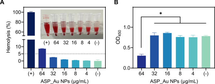
In an effort to simulate in vivo security and additional discover the therapeutic results, we performed hemolysis and cytotoxicity assays. The hemolysis assay, depicted in Fig. 5A, demonstrated that ASP_Au NPs at concentrations 2–8 occasions the MIC (≤ 32 µg/mL) didn’t manifest any hemolytic exercise, remaining under the outlined cutoff of hemolysis charges ≤ 5%. For the cytotoxicity assay, the CCK-8 assay methodology was employed. As illustrated in Fig. 5B, publicity to ASP_Au NPs at concentrations 2–8 occasions the MIC (≤ 32 µg/mL) had no impression on cell viability. Collectively, ASP_Au NPs exhibited antimicrobial and antibiofilm results with out eliciting corresponding toxicity.
In vivo antimicrobial efficiency research
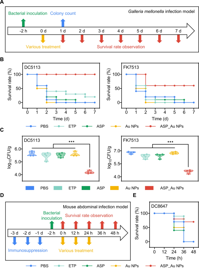
Constructing upon the outstanding in vitro antimicrobial efficacy and strong security profile of ASP_Au NPs, we proceeded to research their results inside a Galleria mellonella an infection mannequin and a mouse acute intra-abdominal an infection mannequin. The Galleria mellonella an infection mannequin was chosen to validate the in vivo antimicrobial efficiency of ASP_Au NPs. A number of authoritative research have highlighted Galleria mellonella‘s position as a secure in vivo mannequin, characterised by its cost-effectiveness, user-friendliness, and absence of moral limitations. This mannequin produces outcomes much like these achieved by vertebrate in vivo experiments [63,64,65]. In our preliminary step, we launched varied concentrations of ASP_Au NPs into wholesome Galleria mellonella to gauge in vivo security and dismiss the potential of drug-induced mortality. Illustrated in Further file 1: Determine S8, all Galleria mellonella (10 per group) injected with ASP_Au NPs survived throughout the huge focus vary examined. Consequently, DC5113 and FK7513 had been chosen because the experimental strains to determine a CRE an infection mannequin. Changes had been made to the strategies in accordance with prior research [66]. The final experimental process is printed in Fig. 6A. Succinctly, wholesome Galleria mellonella weighing between 200–300 mg had been distributed into 5 teams: PBS, ETP, ASP, Au NPs, and ASP_Au NPs, with 10 larvae per group. Bacterial suspensions (DC5113, 10 µL, 1.5 × 10⁸ CFU/mL; FK7513, 10 µL, 7.5 × 10⁶ CFU/mL) had been microsyringe-injected into the penultimate left limb beneath constant circumstances. After a 2 h bacterial publicity, ASP_Au NPs had been injected into the penultimate proper limb at a focus of 16 µg/mL, as decided from the MIC within the in vitro antimicrobial assay. The concentrations of ETP, ASP, and Au NPs remained as beforehand indicated. Larval survival was documented every day, with mortality ascertained within the absence of response to bodily stimuli. As demonstrated in Fig. 6B, Galleria mellonella contaminated with DC5113 and handled with ASP_Au NPs exhibited a 100% survival charge, whereas these contaminated with FK7513 and handled with ASP_Au NPs confirmed a 70% survival charge. Conversely, larvae within the PBS, ETP, ASP, and Au NPs teams predominantly perished inside 7 days. These findings underscored the notable enhancement within the survival charge of CRE-infected Galleria mellonella by ASP_Au NPs, aligning with mouse mannequin outcomes. Moreover, bacterial colony counting was performed to replicate the in vivo antimicrobial results of ASP_Au NPs, with slight decorations based mostly on bacterial pressure traits [67]. Briefly, micro organism proof against CRE strains had been selectively remoted from Galleria mellonella a day after customary therapy. Following the euthanasia of 4 larvae per group, their our bodies had been processed, yielding a suspension in PBS. A ten µL aliquot of the suspension was positioned onto an Luria Bertani (LB) agar plate containing ETP, and visual colonies had been tallied the next day. As depicted in Fig. 6C, the bacterial load in Galleria mellonella handled with ASP_Au NPs was markedly decrease than that within the PBS, ETP, ASP, and Au NPs teams, for each DC5113 and FK7513 infections. The ASP_Au NPs therapy group witnessed practically a 2 log10 CFU/g discount in bacterial load, whereas no vital variations had been famous within the different teams. These outcomes affirm the outstanding in vivo antimicrobial properties of ASP_Au NPs.
In vivo antimicrobial efficiency of ASP_Au NPs. A Basic experimental process of the Galleria mellonella bacterial an infection mannequin. B Survival of Galleria mellonella larvae contaminated with scientific CRE and handled with totally different formulations at totally different time factors. C Bacterial load in Galleria mellonella larvae contaminated with scientific CRE and handled with totally different formulations after 24 h. D Basic experimental process of the acute intraperitoneal an infection mannequin in mice. E Survival of mice contaminated with scientific CRE and handled with totally different formulations at totally different time factors
Relating to the mouse acute intra-abdominal an infection mannequin, Fig. 6D outlines the great process for in vivo mouse experiments. We employed immunosuppressant cyclophosphamide (150 mg/kg) to induce immune-deficient mice, simulating people with compromised immune operate. DC8647 was chosen because the experimental pressure. Following three consecutive intraperitoneal immunosuppressant injections, mice had been inoculated with a bacterial suspension (200 µL, 1.5 × 10⁸ CFU/mL) supplemented with 5% yeast extract into the peritoneal cavity [68]. Given the frequent scientific software of carbapenems like ETP, an ETP management group was integrated. The mice had been divided into 5 teams—PBS, ETP, ASP, Au NPs, and ASP_Au NPs—every consisting of 10 mice. Earlier experiences knowledgeable the strategies employed to assemble mouse fashions in vivo [69]. After a 2 h bacterial publicity, the corresponding medicine had been administered at 0 h, 12 h, and 24 h. ASP_Au NPs had been administered at a focus of 10 mg/kg, as beforehand described, by a 200 µL injection. Mouse survival was monitored each 12 h, alongside measurement of the mice’s physique weight. As revealed in Fig. 6E, mice handled with ASP_Au NPs confirmed a 70% survival charge, whereas these within the PBS, ETP, ASP, and Au NPs teams succumbed inside 48 h. These outcomes spotlight the potential of ASP_Au NPs in countering CRE infections in vivo and considerably bettering the survival charges of contaminated mice. Furthermore, adjustments in mouse physique weight had been tracked as an oblique indicator of the drug’s results. As depicted in Further file 1: Determine S9, mice handled with ASP_Au NPs exhibited a considerably slower lower in physique weight in comparison with these within the PBS, ETP, ASP, and Au NPs teams, underscoring the in vivo anti-infective results of ASP_Au NPs. To conclude, each the mouse mannequin and Galleria mellonella mannequin corroborate the potential of ASP_Au NPs as a promising choice towards scientific CRE infections.
In vivo biocompatibility evaluation
In mild of the demonstrated efficacy of ASP_Au NPs therapy in mice, an in-depth exploration of the inflammatory response and physiological alterations in mice was performed to evaluate the physiological ramifications of ASP_Au NPs in vivo. The mouse mannequin methodology was replicated, aside from the administration of immunosuppressants and bacterial inoculation, with the target of observing the impression of ASP_Au NPs on mouse immune response and physiological capabilities. Initially, histopathological sections had been ready from the guts, liver, spleen, lung, and kidney of mice that had been sacrificed 48 h post-treatment. Hematoxylin and eosin (H&E) staining was carried out to guage the infiltration of irritation in varied organ tissues. As illustrated in Further file 1: Determine S10, the organ tissue morphology in mice handled with ASP_Au NPs appeared regular and much like the PBS group, with no marked indications of irritation infiltration or tissue congestion. This implies that ASP_Au NPs are nonhazardous to mouse organ tissues and don’t elicit extreme inflammatory reactions.
Subsequently, blood samples had been obtained through retro-orbital bleeding for complete blood rely evaluation with a view to monitor the differential leukocyte rely. The outcomes, as proven in Further file 1: Determine S11, indicated that the counts of white blood cells (WBC) and neutrophils (NEUT) in mice handled with ASP_Au NPs had been corresponding to these within the PBS group, with all values falling inside the regular reference vary (WBC: 0.8–10.6 109/L; NEUT: 0.23–3.6 109/L). This implies that ASP_Au NPs didn’t result in a big rise in white blood cell rely that would set off an undue immune response. Moreover, an evaluation of biochemical markers was carried out by subjecting the blood samples to centrifugation and serum testing. This evaluation targeted on eight liver operate markers, 4 cardiac enzyme markers, and three kidney operate markers, all consistent with scientific apply. As depicted in Further file 1: Determine S12, all of the biochemical markers in mice handled with ASP_Au NPs confirmed no notable disparities compared to the PBS group, except for alkaline phosphatase (ALP) and creatinine (CREA) ranges, which had been under the reference vary. In the meantime, the opposite biochemical markers remained inside the regular reference vary, which is printed in Further file 1: Desk S5. It is very important be aware that elevated ALP and CREA ranges would possibly indicate impaired liver or kidney operate. Nevertheless, it normally holds no scientific significance when these markers fall under the reference vary and could be attributed to methodological elements. In conclusion, ASP_Au NPs have demonstrated excellent biocompatibility in vivo, positioning them as promising candidates for potential in vivo purposes.
Analysis of anti-inflammatory results
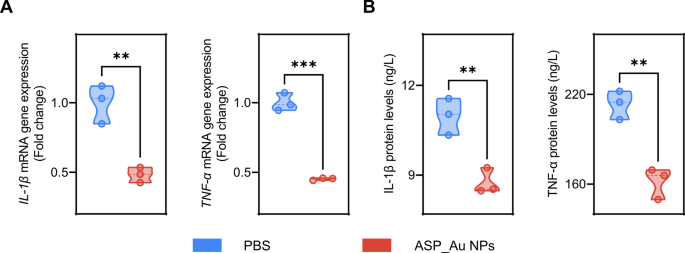
Commercially obtainable NAS has proven particular anti-inflammatory results [70]. As a NAS member, ASP additionally reveals analgesic properties much like nonsteroidal anti-inflammatory medicine, and it might diminish the degrees of the inflammatory cytokine IL-6 [71]. Therefore, the speculation was formulated that ASP_Au NPs may possess potential anti-inflammatory results. To validate this speculation, actual time quantitative PCR (RT-qPCR) and enzyme-linked immunosorbent assay (ELISA) had been performed. For the RT-qPCR experiment, an inflammatory response was triggered in RAW264.7 mouse macrophage cells in vitro using lipopolysaccharide (LPS) [72]. The LPS-stimulated cells had been divided into two teams: the PBS group and the ASP_Au NPs group. The ASP_Au NPs focus was decided based mostly on the MIC from the antimicrobial assay, which was 16 µg/mL. Following 4 h of drug therapy, mobile RNA was extracted for RT-qPCR evaluation to quantify the relative expression ranges of inflammatory cytokines. As depicted in Fig. 7A, the degrees of IL-1β and TNF-α within the ASP_Au NPs group had been considerably downregulated in comparison with the PBS group, indicating the potential anti-inflammatory impact of ASP_Au NPs. For the ELISA experiment, an analogous method to the RT-qPCR experiment was adopted, whereby IL-1β and TNF-α protein detection kits had been utilized to measure the cytokine ranges within the cell supernatant obtained after centrifugation. As proven in Fig. 7B, the protein ranges of IL-1β and TNF-α within the ASP_Au NPs group had been considerably diminished compared to the PBS group, aligning with the outcomes of the RT-qPCR evaluation. Thus, our findings recommend that ASP_Au NPs could maintain potential anti-inflammatory results.


