Precept of detection by the UCNPs-Cas14a-based PEC sensor
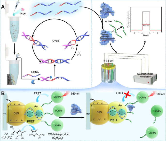
A easy sign conversion methodology was launched through the use of UCNPs-Cas14a-based PEC, with a purpose to lengthen the kinds of analytes that may be detected by CRISPR-based sensors. On the similar time, the sensor achieved photoelectric sign output by the ingenious mixture of CRISPR and nanomaterials, thereby additional enhancing the sensitivity of detection. The working precept of the UCNPs-Cas14a-based PEC sensor is especially divided into three levels (Fig. 1): 1) sign identification and conversion relying on the magnetic bead ATP-cDNA complicated; 2) a lot of goal chains is obtained by SDA for sign amplification; 3) sign output impacts Cas14a-sgDNA the cleavage exercise of which will be activated by these goal DNA. The PEC workstation consists of the working electrode UCNPs-ssDNA-CdS@Au-modified ITO, the counter electrode which is a platinum sheet, and the reference electrode which is Ag/AgCl. A non-specific ssDNA reporter is included into the PEC workstation, which possesses UCNPs for absorbing NIR gentle (980 nm) and transferring this power to CdS@Au/ITO; the sulfhydryl group tethered to the floor of the sensor thereby leads to the era {of electrical} alerts (Fig. 1B). Within the presence of the goal, cDNA dissociates from the aptamer-binding area as a result of the aptamer on the magnetic beads binds to the goal. Free cDNA collected by magnetic separation induces SDA (whereby cDNA and template strand bind to and polymerize alongside the 5’-3’ route with the assistance of polymerase, after which Nt.BsmAI acknowledges and cleaves one of many double strands to kind a niche, in order that KF can enter and polymerize to finish the double strands, and the cleaved fragments are changed, persevering with on this manner for a number of cycles). This course of produces a considerable amount of ssDNA, which triggers the cleavage exercise of the Cas14a-sgRNA complicated, cleaving UCNPs from the floor of the CdS@Au/ITO electrode. With growing distance between UCNPs and CdS@Au/ITO, it turns into more and more troublesome to switch the NIR gentle absorbed by UCNPs to CdS@Au/ITO, so {the electrical} alerts change into weaker. Within the absence of the goal, the cleavage exercise of Cas14a-sgRNA is silenced, so the construction of UCNPs-ssDNA-CdS@Au/ITO stays intact. On the premise of this technique, a common UCNPs-Cas14a-based PEC sensor was developed for sign transduction in a easy and environment friendly method, as reported on this research.
Feasibility and characterization of the UCNPs-Cas14a-based PEC sensor
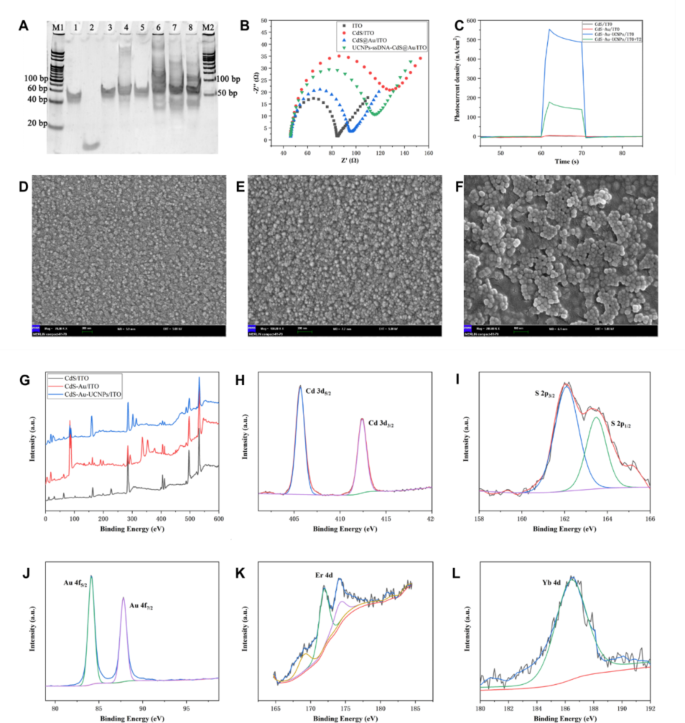
T2 was chosen for evaluating the efficiency of the UCNPs-Cas14a-based PEC sensor for detecting the goal of alternative. The T-2 toxin is likely one of the most potent naturally-occurring sort A mucorene toxins secreted by fusarium, which is extensively current in meals reminiscent of corn and wheat [33, 34]. T-2 is poisonous in hint quantities, and causes injury to a number of organs such because the liver, mind and reproductive system after getting into the human physique [35,36,37]. Subsequently, it’s of nice significance to develop an ultrasensitive and highly-stable technique for detecting and quantifying T2. The important thing to the profitable detection of T2 by the UCNPs-Cas14a-based PEC sensor is that a considerable amount of ssDNA obtained after SDA on the complementary strand comparable to the T2 aptamer can bind to sgRNA, thereby triggering the cleavage exercise of Cas14a and inflicting modifications within the photocurrent. First, the complementary strand [38], template strand and corresponding sgRNA sequence had been designed based mostly on the T2 aptamer. The impact of cDNA-induced SDA was verified by polyacrylamide gel electrophoresis (PAGE). As proven in Fig. 2A, within the presence of solely T-DNA within the system, no new bands had been present in Lane 1, indicating that no amplification had occurred. Within the presence of each complementary strand and T-DNA however with out enzyme (Lane 3/4/5), no new bands had been discovered both, however the authentic band at 50 bp was shifted barely upward, confirming pairing between the complementary strand and T-DNA. Within the presence of the complementary strand, T-DNA and enzymes, new bands appeared in Lane 6/7/8 at about 30 bp, related in size to the goal ssDNA, indicating that SDA had been profitable and the goal product generated. Furthermore, with growing cDNA focus, the colour of the brand new bands grew to become deeper. These outcomes exhibit that cDNA can particularly induce SDA, and that the quantity of ssDNA SDA is immediately proportional to the cDNA focus. As well as, to substantiate the flexibility of ssDNA SDA to activate Cas14a, PEC research had been additional carried out. It might be noticed underneath a scanning electron microscope (Fig. 2D, E and F) that the electrode was efficiently modified with CdS, Au and UCNPs. Alternating-current impedance spectroscopy is a extremely efficient methodology to explain the resistance of floor modified electrodes, and the diameter of the semicircle within the impedance spectrum corresponds to the worth of resistance (Ret) of electron switch. By evaluating the curves in Fig. 2B, it may be seen that after modification with Au-NPs, the ITO Ret barely declined, which was associated to the superb conductivity of Au. After modification with CdS QDs, the ITO Ret significantly elevated, the reason is that the quantum dot (QD) itself is a semiconductor with poor conductivity. After deposition of Au nanoparticles on the floor of CdS QDs, the modified electrode Ret declined however was nonetheless larger than that of the electrode modified by Au alone. Furthermore, the UCNPs-ssDNA-CdS@Au/ITO electrode Ret was larger than that of CdS@Au/ITO, as a result of the conductivity of UCNPs can be poor. Nevertheless, its Ret was decrease than that of CdS/ITO, which can even be attributed to the superb conductivity of Au. Within the absence of T2, the photoelectric sign noticed was the strongest, as a result of Cas14a isn’t activated and upconversion happens near the electrode floor (Fig. 2C). Within the presence of T2, the photoelectric sign grew to become weaker, inversely proportional to the T2 focus. This means that the cleavage exercise of Cas14a had been triggered, in order that the upconversion happens distant, thus hindering power switch. The complete XPS spectra of the modified electrode in every step are proven in Fig. 2G-M. Extra characterization of the working electrode UCNPs-ssDNA-CdS@Au/ITO and photoelectric sign conversion course of evaluation was proven within the supporting info.
Feasibility and electrode characterization of UCNPs-Cas14a based mostly PEC. A) PAGE electropherograms to confirm complementary strand triggered SDA of T2 aptamers (M1: 20 bp marker; 1: T-DNA; 2: cDNA; 3: cDNA + T-DNA; 4: cDNA + T-DNA + Nt; 6/7/8: 0.5/2/5 µM cDNA + T-DNA + KF + Nt; M2: 50 bp marker). DNA + KF; 5: cDNA + T-DNA + Nt; 6/7/8: 0.5/2/5 µM cDNA + T-DNA + KF + Nt; M2: 50 bp marker). B) AC impedance spectrum of the modified electrode [5 Mm Fe(CN)63-/4-(1:1), 0.1 M KCl, 100 KHZ-0.1 HZ]. C) Feasibility validation of UCNPs-Cas14a based mostly PEC. D) SEM of CdS/ITO. E) SEM of CdS@Au/ITO. F) SEM of UCNPs-ssDNA-CdS@Au/ITO. G) XPS spectra of modified electrodes. XPS spectra of modified electrodes of Cd (H), S (I), Er (J), Au (Okay) and Yb (M)
Optimization of the detection efficiency of the UCNPs-Cas14a-based PEC sensor
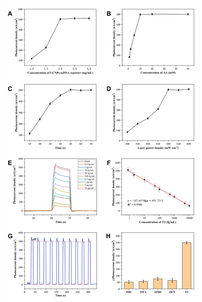
The cleavage effectivity of Cas14a protein and conversion effectivity of photoelectric alerts are extraordinarily vital components influencing the detection efficiency of UCNPs-Cas14a-based PEC sensors. As a way to obtain an environment friendly floor chemistry-based trans-cleavage, accessibility of Cas14a to the nonspecific ssDNA is essential. Subsequently, the affect of the density of the UCNPs-ssDNA reporter on the electrode floor on the modifications in photoelectric alerts earlier than and after cleavage was optimized on this research. As will be seen from Fig. 3A, the perfect focus of the UCNPs-ssDNA reporter was 2 mg/mL, creating sufficient area for cleavage of Cas14a on the electrode floor. Excessive-concentration ssDNA reporter molecules will cut back the sign change worth, as a result of such excessive floor density will result in a steric-hindrance impact, thereby limiting cleavage exercise. Furthermore, the effectivity of conversion of photoelectric alerts is affected by the AA focus within the electrolyte, by the cleavage time and by the depth of irradiation by the laser. As proven in Fig. 3B, C and D, with growing AA focus, cleavage time and laser depth, the photocurrent was regularly enhanced till a plateau was reached. Contemplating value financial savings and guaranteeing photoelectric conversion effectivity, the optimum detection circumstances had been decided. These had been as follows: AA focus 10 mM, cleavage time 50 min and laser depth 500 mW·cm− 2.
UCNPs-Cas14a based mostly PEC assay for T2. A) Optimization of UCNPs-ssDNA focus. B) Optimization of AA focus. C) Optimization of response time. D) Optimization of laser depth. E) I-T bar curves comparable to totally different concentrations of T2. F) Normal curves of UCNPs-Cas14a based mostly PEC for T2 detection. G) Stability of UCNPs-Cas14a based mostly PEC sensing. H) Specificity of UCNPs-Cas14a based mostly PEC sensing
T2 detection utilizing the PEC sensor
To find out whether or not the UCNPs-Cas14a-based PEC sensor can detect T2 in a quantitative method, the photocurrent comparable to totally different concentrations of T2 was first measured in a separate system. Sign identification, conversion and amplification experiments had been carried out in an EP tube, and the photocurrent was measured in a detection cell fabricated in-house. It was discovered that the photocurrent was enhanced with growing T2 focus from 0.8 fg/mL to 10,000 fg/mL (Fig. 3E). There was an excellent linear relationship between photocurrent density and the logarithm of T2 focus (Fig. 3F). The regression equation was y = -107.4474lgx + 491.7213 (fg/mL, R2 = 0.9958, n = 8). In accordance with m+-3σ (the place σ is commonplace deviation of the clean, and m is the imply of 10 clean samples), the restrict of detection was estimated to be 0.5128 fg/mL. The I-T columnar curve was plotted underneath a periodic cycle of sunshine “on” and “off” to analyze the steadiness of UCNPs-ssDNA-CdS@Au/ITO in detecting T2 (Fig. 3G). The outcomes confirmed that the relative commonplace deviation (RSD) of the photoelectric response worth to T2 was 0.68% underneath 10 cycles of sunshine “on” and “off”, indicating that the electrode had good stability.
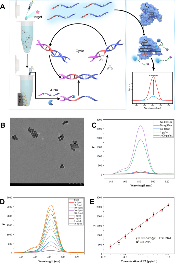
On the similar time, a Cas14a-based upconversion fluorescence (UCNPs-Cas14a-based FL) sensor was additionally constructed to detect T2 as a comparability (Fig. 4A). UCNPs had been characterised by TEM, displaying that that they had a spherical form with a particle dimension of about 30 nm (Fig. 4B). The outcomes proven in Fig. 4C indicated that fluorescence was considerably generated solely within the presence of the goal. The optimization was proven in Determine S4. The sign identification, conversion and amplification properties of the UCNPs-Cas14a-based FL sensor had been the identical as these for the UCNPs-Cas14a-based PEC sensor, however with a fluorescence sign output. In different phrases, the mixture of SDA product ssDNA and Cas14a-sgRNA complicated can activate the ssDNA of the UCNPs-ssDNA-D probe within the Cas14a trans-cleavage system. As the gap between UCNPs and Dabcyl grew to become bigger, the fluorescence of UCNPs at 482 nm recovered. It might be seen from Fig. 4D and E that the T2 focus was immediately proportional to the fluorescence depth. When the T2 focus was inside 0.02–10 pg/mL, the linear equation was y = 825.5435lgx + 1791.2164 (pg/mL, R2 = 0.9915, n = 8, LOD = 0.0133 pg/mL). In contrast with UCNPs-Cas14a-based FL sensors, the sensitivity of the UCNPs-Cas14a-based PEC sensor was improved about 25-fold, whereas its stability was not inferior. Subsequently, it’s concluded that the excessive sensitivity of UCNPs-Cas14a-based PEC sensors advantages from the triple amplification of SDA, Cas14a trans-cleavage characteristic and photoelectric alerts.
UCNPs-Cas14a based mostly FL detection of T2. A) Schematic illustration of UCNPs-Cas14a based mostly FL detection. B) TEM of UCNPs, C) Feasibility of fluorescence detection. D) Fluorescence spectra comparable to totally different concentrations of T2. E) Normal curve of UCNPs-Cas14a based mostly FL for T2 detection
Analysis of detection efficiency of PEC sensor when it comes to specificity and measurement of T2 in oats
In view of the detection efficiency of the UCNPs-Cas14a-based PEC sensor underneath experimental circumstances, it’s essential to substantiate its specificity and sensible utility for goal detection. To this finish, the specificity of the UCNPs-Cas14a-based PEC sensor was examined utilizing analogues of the T2 toxin, reminiscent of zearalenone (ZEN), ochratoxin A (OTA), fumonisin (FB1) and aflatoxin B1 (AFB1). As proven in Fig. 3H, even excessive concentrations of those analogues barely induced any modifications within the generated photocurrent in contrast with the background/clean alerts. In distinction, T2 toxin at a focus 100 instances decrease than the analogues nonetheless induced clearly measurable modifications within the photocurrent. Subsequently, the UCNPs-Cas14a-based PEC sensor can be utilized for extremely particular T2-targeted detection.
Subsequent, oats had been chosen as a consultant of precise real-world samples and T2 detected by the UCNPs-Cas14a-based PEC sensor was assessed. Methodology comparability was additionally carried out with a industrial equipment (restrict of detection 15 pg/mL). The ELISA equipment outcomes had been in good settlement with the UCNPs-Cas14a-based PEC sensor. The ensuing evaluation in Desk S1 revealed that the standard-addition restoration charge of T2 in oats was 84.49–118.30%, and its RSD was 1.4–2.9%, suggesting that the UCNPs-Cas14a-based PEC sensor can function an elective methodology for quantitative detection of T2 in oats.
Detection of protein tyrosine kinase 7 (PTK7) used PEC sensor
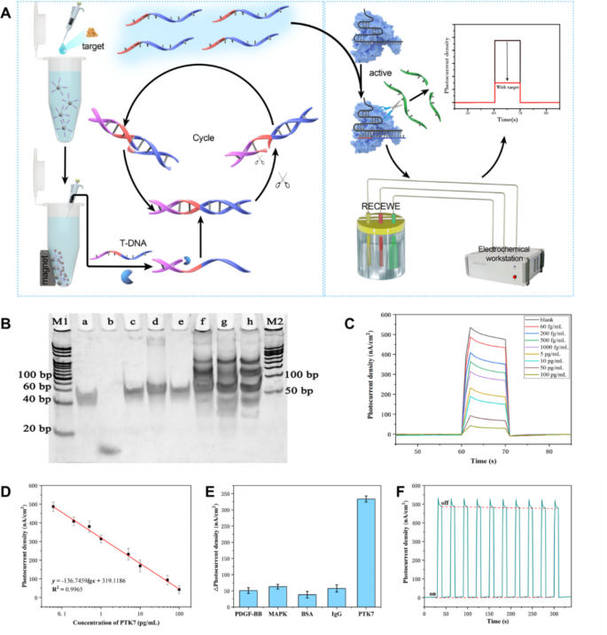
To research the final applicability of the UCNPs-Cas14a-based PEC sensor, its skill to detect protein targets was explored. PTK7 is a kinase that catalyzes the switch of γ-phosphate from ATP to protein tyrosine residues. It performs an vital position in cell progress, proliferation and differentiation [39, 40]. Most PTKs found to this point are oncogene merchandise of oncogenic RNA viruses, they usually may also be produced by proto-oncogenes in vertebrates [41]. Subsequently, they’re vital tumor markers within the clinic [42]. Right here, PTK7 was used because the goal, and the aptamer served as the popularity component of PTK7. The precept for its detection is proven in Fig. 5A. Within the presence of PTK7, the corresponding cDNA was dissociated and might be collected by magnetic separation. cDNA might induce SDA to acquire a considerable amount of ssDNA, thereby triggering the cleavage exercise of the Cas14a-sgRNA complicated, and resulting in cleavage of UCNPs from the floor of the CdS@Au/ITO electrode. Because of this, power switch was hindered, weakening {the electrical} alerts. The gel electrophoretogram (Fig. 5B) confirmed that the complementary strand might efficiently induce SDA, that’s, new bands appeared at about 30 bp within the Lane f/g/h. There have been no new bands (Lane a/b/c/d/e) when both the T-DNA, or enzyme, or cDNA was absent.
UCNPs-Cas14a based mostly PEC for PTK7 detection. A) Schematic illustration of UCNPs-Cas14a based mostly PEC for PTK7 detection. B) PAGE electropherogram verifying the SDA triggered by the complementary DNA of PTK7 aptamer (M1: 20 bp marker; a: T-DNA; b: cDNA; c: cDNA + T-DNA; d: cDNA + T-DNA + KF; e: cDNA + T-DNA + Nt; f/g/h: 0.5/2/5 µM cDNA + T-DNA + KF + Nt; M2: 50 bp marker). C) I-T bar curves comparable to totally different concentrations of PTK7. D) Normal curve of UCNPs-Cas14a based mostly PEC for the detection of T2. E) Specificity of UCNPs-Cas14a based mostly PEC sensing. F) Stability of UCNPs-Cas14a based mostly PEC sensing to detect PTK7
Accordingly, detection of PTK7 utilizing the UCNPs-Cas14a-based PEC sensor was assessed. As proven in Fig. 5C and D, there was an excellent linear relationship between photocurrent density and the logarithm of PTK7 focus when the latter was inside the vary 0.06–100 pg/mL. The regression equation was y = -136.7459lgx + 319.1186 (pg/mL, R2 = 0.9977, n = 8). The calculated restrict of detection was 0.03783 pg/mL.
The UCNPs-Cas14a-based PEC sensor confirmed higher selectivity for PTK7 at a focus of 10 pg/mL than for the opposite competing proteins examined (platelet-derived progress factor-BB, mitogen-activated protein kinase, BSA and IgG) (Fig. 5E). It may be seen that the aptamer employed has wonderful specificity within the sensor. The analysis of stability was proven in Fig. 5F. All baseline and photocurrent responses (n = 10) had been comparatively secure in “on” and “off” states, respectively.
Moreover, the flexibility of the UCNPs-Cas14a-based PEC sensor to detect PTK7 in human serum samples was additionally examined. The outcomes confirmed (Desk S2) that the standard-addition restoration charge in human serum was 83.34–118.40%, and its RSD was lower than 4%, confirming the reliability of detection by this UCNPs-Cas14a-based PEC sensor. Furthermore, the aptamer is a common recognition component for proteins and small molecules. Subsequently, the UCNPs-Cas14a-based PEC sensor will be utilized to a wide range of analytes.


