Preparation and characterization of PVP-Ir NPs and composite fiber movies
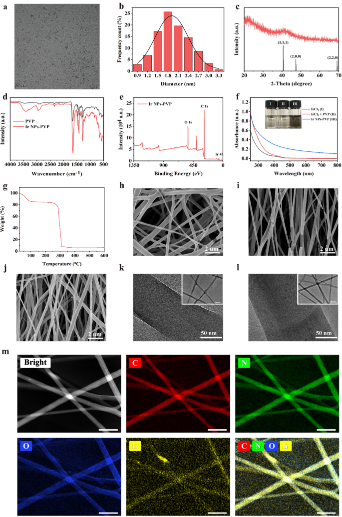
In experiments, PVP-Ir NPs had been artificial primarily based on the earlier studies with minor modifications. Briefly, the precursor iridium trichloride hydrate was first dissolved in H2O and handled by ultrasound till a clarified resolution was obtained. Then the answer was added into the PVP contained ethanol solvent and stirred in a single day at room temperature. After that, a vivid yellow resolution was acquired after which refluxed for an additional 8 h at 100 ℃. The ensuing brown resolution was handled by rotary evaporation to take away the solvent, and black manufacturing was obtained on the finish. Transmission electron microscopy (TEM) demonstrated that the PVP-Ir NPs confirmed a subspherical construction, and no obvious aggregation was noticed (Fig. 1a). In the meantime, the statistical evaluation of 300 nanoparticles primarily based on TEM pictures confirmed that the majority PVP-Ir NPs distributed at 1 to three nm diameter with a median measurement of roughly 1.8 nm (Fig. 1b). The diffraction peaks at 40.66°, 47.32°, and 69.14° had been sharp and intense primarily based on the outcomes of X-ray diffraction (XRD) evaluation, indicating the crystal-like construction of the particles (Fig. 1c), which is in keeping with the JCPDS NO. 06–0598. The profitable floor coating by PVP was confirmed by the Fourier rework infrared spectra (FT-IR) (Fig. 1d), which have a attribute absorbance peak at 1286 and 1657 cm−1 for C–N and C=O bonds. Moreover, X-ray photoelectron spectroscopy (XPS) recognized the digital state of Ir(0), which contributed to the catalytic mechanism of iridium nanozymes. And efficiently coordinated pyrrole teams of PVP to nanozymes was additionally confirmed primarily based on the outcomes of XPS (Fig. 1e). Moreover, the UV–vis spectrum and the corresponding shade modifications of the answer verified the fabrication of nanozymes (Fig. 1f). Moreover, the Thermogravimetric evaluation (TGA) additional demonstrated the PVP exiting, with an virtually 80% weight ratio in PVP-Ir NPs (Fig. 1g).
a TEM outcomes of synthesized nanozymes. b Measurement distribution evaluation of 300 random nanoparticles. c XRD outcomes of nanozymes. d FTIR evaluation of PVP and PVP coated nanozymes. e XPS outcomes of nanozymes. f UV spectra evaluation of varied options. The insert exhibits the photographs of the IrCl3 (I), IrCl3 + PVP (II), PVP-Ir NPs (III). g TG evaluation of PVP-Ir NPs. SEM picture of h GelMA/R, i GelMA/A, j Ir@GelMA/A. TEM pictures of ok GelMA fibers, and l Ir@GelMA fibers. m EDS evaluation of PVP-Ir NPs loaded GelMA fibers. The dimensions bars are 20 nm in a, 2 μm in h, i, and j, 50 nm in ok, l, and m, and 500 nm within the insert pictures
To acquire the hydrogel fiber movie with fascinating microstructure and multi-functions, photocrosslinkable GelMA was electrospun with a rotating collector with variable speeds. The obtained GelMA fibrous movies with random or aligned fibers association had been named GelMA/R and GelMA/A, respectively, whereas the one with aligned fibers and loaded PVP-Ir NPs was named Ir@GelMA/A. Scan transmission electron microscope (SEM) indicated that the random and aligned crosslinked microstructure of hydrogel fiber movies could possibly be fashioned below totally different collector rotation speeds (Fig. 1h–j). Primarily based on the SEM outcomes, the diameters of random fibers had been distributed between 250 and 450 nm (Extra file 1: Fig. S1a). The excessive collector rotating pace of about 2500 rpm/min may cut back the diameter of fibers within the aligned teams, much like the earlier literature (Extra file 1: Fig. S1b). Nevertheless, the doping of PVP-Ir NPs into the aligned fibers below the high-speed situation elevated the typical diameter of fibers (Extra file 1: Fig. S1c). TEM and EDS evaluation demonstrated the profitable encapsulation of PVP-Ir NPs (Fig. 1ok–m). The GelMA fibers confirmed a clear inside with out nanoparticles loaded (Fig. 1ok). The black dots, nevertheless, had been distributed within the fibers after electrospinning (Fig. 1l). The EDS evaluation confirmed the homogeneous distribution of Ir components within the fibers (Fig. 1m), referring to the profitable encapsulation of PVP-Ir NPs. Moreover, the outcomes of ICP-MS demonstrated the concentration-dependent loading means of PVP-Ir NPs (Extra file 1: Fig. S2).
Electrospun fiber movie loaded with iridium nanozymes simulates a number of antioxidant enzymes in vitro
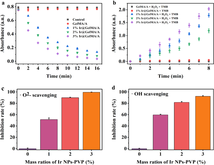
To research the aptitude of ROS scavenging, a chains of enzyme exercise experiments had been utilized to certify the multi-enzymes mimicking impact of varied concentrations of PVP-Ir NPs loaded within the aligned GelMA hydrogel fiber movies, specifically 1%, 2%, and three% Ir@GelMA/A, respectively. First, the CAT-like exercise was examined by decreasing H2O2 via the UV absorption spectrum at 240 nm. The outcomes indicated a time-dependent and concentration-dependent H2O2 degradation by the Ir@GelMA/A teams, whereas no obvious modifications had been noticed between the Management and pure GelMA/A teams, as proven in Fig. 2a. Subsequent, the business reagent Catalase was set as a optimistic management, demonstrating the CAT-like exercise of Ir@GelMA/A (Extra file 1: Fig. S3). Contemplating the manufacturing of oxygen from H2O2 below the catalysis of CAT, the technology of bubbles and the floating of movies additional demonstrated the CAT-like of Ir@GelMA. In distinction, pure GelMA/A movie stayed on the backside in the course of the experiments (Extra file 1: Fig. S4). To instantly verify the manufacturing of oxygen below the catalysis of CAT-like exercise, we carried out a dissolved oxygen check to make clear the generated oxygen. The outcomes confirmed that the focus of the oxygen elevated because the catalysis proceeded, whereas no obvious modifications had been noticed within the management and the pure fiber movie teams (Extra file 1: Fig. S5). To discover the POD mimicking exercise of Ir@GelMA/A, the three,3′,5,5′-tetramethylbenzidine (TMB) was utilized to guage the catalysis impact. The numerous time-dependent and concentration-dependent modifications of the absorption values of TMB-ox at 652 nm confirmed the POD-like of the fiber movies encapsulated with PVP-Ir NPs. Nevertheless, the absorption worth of TMB-ox was practically unchanged with out H2O2 or PVP-Ir NPs loading, respectively (Fig. 2b).
a CAT-like exercise of varied mass ratios of Ir@GelMA/A (n = 3 for every group). b POD-like exercise of varied ratios of Ir@GelMA/A (n = 3 for every group). c SOD-like exercise of varied mass ratios of Ir@GelMA/A (n = 3 for every group). d Hydroxyl radicals scavenging impact of varied mass ratios of Ir@GelMA/A (n = 3 for every group)
Superoxide can be the frequent ROS generated in vivo. Thus, the superoxide dismutase equipment was used to detect the SOD mimetic means of fiber movies containing totally different concentrations of the nanozymes. The Ir@GelMA/A fiber movies confirmed a concentration-dependent relationship of superoxide radicals scavenging means with roughly 51, 89, and 99% of the •O2− decomposed by 1%, 2%, and three% PVP-Ir NPs loaded inside 60 min, respectively (Fig. 2c). Furthermore, virtually 92% of hydroxyl radical (•OH) was decomposed by 3% Ir@GelMA/A, which confirmed excessive sensitivity to •OH (Fig. 2d). These outcomes indicated that Ir@GelMA/A possessed multi-enzymes mimetic means towards numerous ROS.
Aligned electrospun fiber movie loaded with iridium nanozymes eliminates intracellular ROS and induces cell orientation development
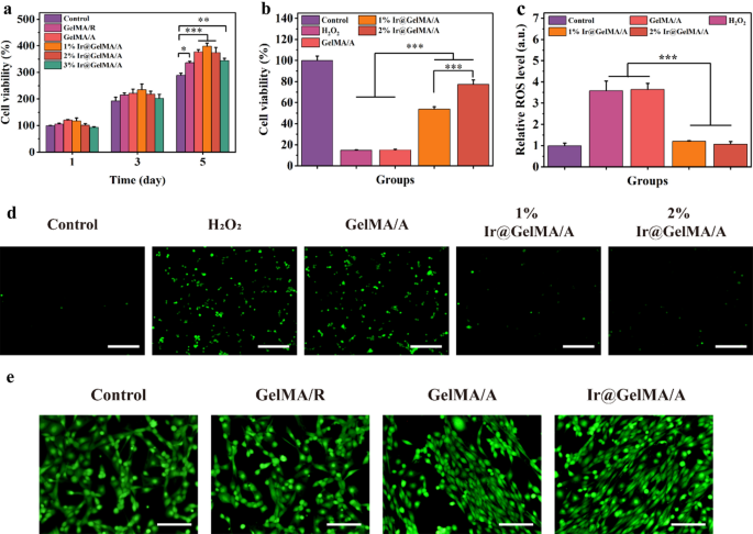
H2O2 and O2− are essentially the most plentiful ROS produced intracellular in the course of the wound course of. Physiologically, cells can generate multifarious antioxidant enzymes stimulated by average oxygen stress to scavenge ROS. Nevertheless, the overwhelming manufacturing of ROS could break this concord and result in cell loss of life. Inspired by the aforementioned traits of Ir@GelMA/A, we evaluated their potential means to scavenge ROS on the mobile stage and cytoprotective impact. First, we examined the cytocompatibility of synthesized fiber movies. As proven in Fig. 3a, NIH-3T3 cells on the GelMA hydrogel fiber movie had higher viability and proliferation than the cells on the tradition plate. These variations had been as a result of good biocompatibility and higher physiological setting supplied by GelMA hydrogel for cell proliferation. Moreover, the oriented construction of fiber movie made the cell adhesion simpler and grew sooner, thus having an improved proliferation within the GelMA/A, 1% GelMA/A, and a pair of% GelMA/A teams than the GelMA/R group. Nevertheless, fiber movie loaded with 3% PVP-Ir NPs barely impeded the viability of cells. Afterward, the antioxidant means of varied fiber movies was analyzed. Contrasted with the Management or GelMA/A teams, the proliferative exercise of the cells was promoted coculturing with 1% and a pair of% Ir@GelMA/A (Fig. 3b), suggesting the numerous protecting means of Ir@GelMA/A towards oxidative stress damage. As well as, the quantification of the mobile ROS by the circulate cytometry experiments licensed the wonderful ROS scavenging exercise of fiber movie loading with PVP-Ir NPs (Fig. 3c). Moreover, the fluorescence micrography indicated an elaborated ROS stage after H2O2 therapy, during which distinguished decreases had been noticed each in 1% and a pair of% Ir@GelMA/A handled teams (Fig. 3d). Then, we assessed the power of aligned fiber movie to induce cell orientation. The obtained fluorescence pictures revealed that cells seeded on GelMA/R grew disorderly. Alternatively, the directional development of cells alongside the nanofibers was noticed on GelMA/A fibrous movie. Furthermore, the power of the GelMA/A fiber movie to induce orientation was not impaired with 2% PVP-Ir NPs loaded (Fig. 3e). And the angle distribution evaluation of cells additional demonstrated the conclusions talked about above (Extra file 1: Fig. S6). Subsequently, the two% Ir@GelMA/A fibrous movie was chosen for additional experiments primarily based on the outcomes talked about above.
a The cell viability of NIH-3T3 after totally different therapies. b The cell protecting impact of PVP-Ir NPs loaded fiber movie towards H2O2 therapy. c The circulate cytometry quantification of mobile ROS stained by DCFH-DA. d Fluorescence photographs of the intracellular ROS. e The impact of inducing cell orientation development of varied GelMA fiber movies. The dimensions bars are 200 μm in d, and e
Composite fiber movie loaded with silver nanoparticles inhibits bacterial development
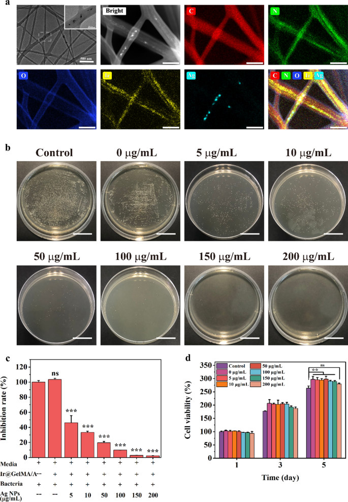
Including the varied concentrations of Ag NPs to the pre-electrospinning resolution, we obtained Ag NPs and PVP-Ir NPs co-loaded within the aligned GelMA fiber movies, named as Ir@Ag@GelMA/A. The big nanoparticles and small dots noticed within the TEM pictures and the evaluation of the corresponding components indicated the simultaneous loading of each nanoparticles (Fig. 4a). To make clear the potential antibacterial exercise of the multifunction GelMA hydrogel fiber movie, gram-positive Staphylococcus aureus (S.aureus) had been chosen to coculture with numerous concentrations of Ag NPs (0–200 μg/mL) loaded fiber movies. The outcomes of bacterial colony depend and quantification evaluation confirmed that the S.aureus within the PBS and Ir@GelMA/A teams maintained regular viability. Moreover, the inhibition charge towards the bacterial was positively related to the loading concentrations of Ag NPs. Ir@GelMA/A fiber movie with about 150 μg/mL Ag NPs loaded may inhibit the expansion of S.aureus utterly (Fig. 4b, c). As well as, we additionally demonstrated the biocompatibility of Ag NPs loaded GelMA fiber movie. The cell-material coculture experiments confirmed good viability and proliferation of NIH-3T3 cells seeded on totally different fiber movies much like that seeded on the plastic plate, whereas with barely compliance to the viability within the 200 μg/mL group (Fig. 4d). Furthermore, the SEM pictures confirmed the addition of such nanoparticles didn’t have an effect on the aligned construction of the fibers within the Ir@Ag@GelMA/A bunch (Extra file 1: Fig. S7), and the correspondingly cell experiments additional demonstrated that the Ir@Ag@GelMA/A can nonetheless markedly induce the orientation development of cells as Ir@GelMA/A did (Extra file 1: Fig. S8). Thus, we chosen the two% PVP-Ir NPs and 150 μg/mL Ag NPs loaded Ir@Ag@GelMA/A for the additional experiments.
a TEM pictures and EDS evaluation of Ir@Ag@GelMA fibers. b The antibacterial impact towards S.aureus of varied concentrations of Ag NPs loaded Ir@Ag@GelMA fibrous movies. c Quantification evaluation of antibacterial impact. d The biocompatibility of varied concentrations of Ag NPs and a pair of% PVP-Ir NPs loaded GelMA fiber movie. The dimensions bars are 200 nm in a, and 200 mm in b
Multifunctional composite fiber movie has admirable water retention means, self-degradation property, and improved mechanical power
A fascinating dressing ought to enhance the alternate of vitamins and metabolites with good water absorbance and retention means for wound therapeutic. Thus, we evaluated the wettability of the GelMA fiber movie. Because the outcomes confirmed, the GelMA/R and GelMA/A fiber movie may take in water for nearly 70 occasions its unique weight, and the water was retained over 48 h. Furthermore, the nanoparticles loaded didn’t impaired the swelling and water retention properties (Fig. 5a). The dressing utilized in wound therapeutic gives non permanent assist and organic results for cell adhesion and proliferation however degrades after activating the tissue development. Thus, the degradation charge of the obtained fiber movie was additional investigated in vitro simulation experiments. Owing to the hydrophilicity nature of GelMA hydrogel, the fiber movie degraded slowly (Fig. 5b). Nevertheless, the Ir@Ag@GelMA/A bunch had the bottom degradation charge, whereas the GelMA/R degraded the quickest. The outcomes demonstrated the biodegradability of the obtained GelMA fibrous movies, which contributed to the biosafety utilized in vivo. After in vivo utility of the hydrogel fiber movie, the capability to withstand deformation is essential for upkeep of structural stability. Subsequently, the tensile check was utilized to testify the mechanical power of varied fiber movie. Outcomes confirmed that every one the GelMA fiber movies exhibit the everyday stress–pressure curve of viscoelastic supplies. Amongst them, GelMA/R had the bottom mechanical power and the longest elongation at break. The aligned construction and the encapsulation of nanoparticles correspondingly elevated the tensile power of the fiber movie (Fig. 5c).
The aligned fiber movies induce elevated cell migration in a wound-healing check
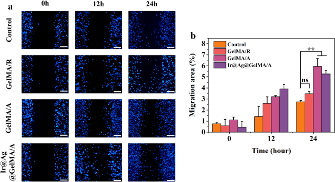
Cell migration performs a pivotal function in lots of complicated physiological and pathological processes, particularly throughout wound therapeutic. In an effort to display the power of the composite fiber movie for selling cell migration and its potential affect on wound therapeutic, we utilized the non-injury culture-insert assay to review cell migration in vitro. In the course of the 24 h experiment interval, the NIH-3T3 cells confirmed continued migration in every group, and the aligned fiber construction clearly improved the migration pace. Furthermore, loading of nanoparticles didn’t impede such capability (Fig. 6).
Ir@Ag@GelMA/A fiber movie promotes the therapeutic technique of experimental contaminated wound fashions
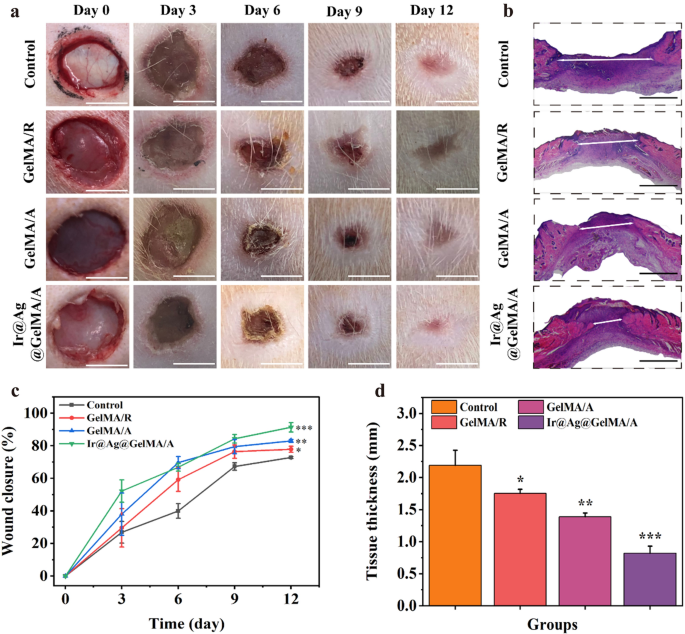
To additional display the useful efficiency of the GelMA fiber movie, we utilized in vivo experiments utilizing contaminated wound fashions (Fig. 7). The rats had been modeled on their again pores and skin and added with bacterial suspension. After the an infection fashions being established, all of the rats had been separated into 4 teams randomly, handled with PBS resolution (Management), GelMA/R, GelMA/A, and Ir@Ag@GelMA/A, respectively. The situations of every wound had been noticed and photographed on days 0, 3, 6, 9, and 12 (Fig. 7a). The wound closure charges had been additionally calculated at each cut-off date in comparison with the unique wound space (Fig. 7c). The outcomes confirmed that the Management group had the bottom therapeutic charge throughout all the experiments, displaying a 72.79 ± 0.60% wound closure charge on day 12. The opposite three teams utilized with GelMA fiber movies confirmed a sooner therapeutic course of than the Management group, with 77.83 ± 1.86%, 82.92 ± 0.95%, and 91.32 ± 2.94% closure charges for GelMA/R, GelMA/A, and Ir@Ag@GelMA/A, respectively. The hydrogel movies’ glorious mechanical service and water retention means supported cell adhesion and development, selling the alternate of vitamins and metabolites, thus accelerating wound therapeutic. In contrast with the GelMA/R group, the aligned construction of fibers within the GelMA/A and Ir@Ag@GelMA/A teams was beforehand verified to information directional cell development and enhance cell proliferation (Fig. 3a, e), displaying a sooner closure charge in vivo experiment. Apart from, ROS manufacturing after wounding will retard the therapeutic course of, and an infection additional exacerbates the wound. The multi-particles doped within the Ir@Ag@GelMA/A bunch may kill micro organism and remove ROS, considerably accelerating wound therapeutic and enhancing wound closure charge. H&E staining of the regeneration pores and skin tissues was utilized to additional confirm the therapeutic outcomes (Fig. 7b). The Ir@Ag@GelMA/A bunch offered the thinnest granulation tissue of 0.82 ± 0.11 mm, whereas the Management group possessed the utmost thickness being 2.19 ± 0.23 mm. The thickness within the GelMA/R and GelMA/A teams was additionally thinner than within the Management group, being 1.75 ± 0.06 mm and 1.39 ± 0.06 mm, respectively (Fig. 7d). These outcomes indicated that encapsulation of PVP-Ir NPs and Ag NPs into GelMA fiber movie would enhance the wound curing.
a Pictures of wounds handled with PBS (Management), GelMA/R, GelMA/A, and Ir@Ag@GelMA/A fiber movies. b Outcomes of H&E staining of Management, GelMA/R, GelMA/A, and Ir@Ag@GelMA/A teams after 12 days. c Wound closure charge of varied teams (n = 3 for every group). d The H&E staining histologic longitudinal sections (n = 3 for every group). The dimensions bars are 5 mm in a, and 1 mm in b
Ir@Ag@GelMA/A fiber movie accelerates the collagen reworking and neovascularization, inhibits irritation, and eliminates the in situ ROS
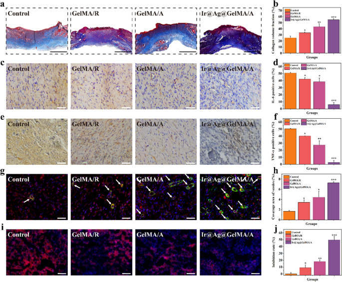
Deposition of collagen on the wound website represents the last word stage within the therapeutic course of. Subsequently, Masson’s trichrome staining measured collagen secretion in all teams (Fig. 8a, b). The cell adhesion and proliferation had been supported within the GelMA/R and GelMA/A teams evaluating with the Management group, selling regular fibroblasts’ viability and secretion exercise. Due to the scavenging ROS capability of PVP-Ir NPs and the antibacterial impact of Ag NPs, offering essentially the most appropriate physiological setting for fibroblasts, essentially the most ample deposition of collagen was noticed within the Ir@Ag@GelMA/A bunch. Extreme irritation attributable to pores and skin harm and an infection slowed down the closure charge in the course of the curing course of. Thus, we detected the expression of the interleukin-6 (IL-6) and tumor necrosis factor-α (TNF-α), utilizing immunohistochemistry experiment on day 12 (Fig. 8c–f). An extra expression of such elements was confirmed within the Management group, demonstrating a extreme irritation. Conversely, each the GelMA/R and GelMA/A teams expressed fewer inflammatory elements than the Management group, primarily benefiting from the mechanical barrier of the movie that stops additional an infection. Furthermore, a big discount of IL-6 and TNF-α was noticed in Ir@Ag@GelMA/A due to the synergistic impact of the loaded nanoparticles.
a Masson’s trichrome staining in numerous teams. b Quantification evaluation of collagen quantity fraction. Immunohistochemical outcomes of c IL-6 and e TNF-α of every group, and the quantification evaluation of IL-6 d, and TNF-α f optimistic cells, respectively. g Double immunofluorescence staining of α-SMA and CD31 for neovascularization. The vessels had been pointed with white arrows. h Quantification evaluation of protection space of recent blood vessels. i Double staining pictures of dihydroethidium (DHE) and DAPI of wounds after totally different therapies. j Quantification evaluation of ROS inhibition charge. The dimensions bars are 1 mm in a, and 200 μm in b–e
Angiogenesis is one other essential indicator for remolded tissues, and the GelMA fiber movies have been demonstrated to advertise tissue vascularization. Thus, we examined the expression of CD31 and α-SMA, which had been the symbols of the endothelial cell and clean muscle cell of the vascular, respectively, by double immunofluorescence to additional confirm the neovascularization (Fig. 8g, h). Outcomes confirmed that only some optimistic staining cells was detected within the Management group, indicating the cells’ impaired migration and differentiation within the extreme inflammatory and an infection state. In distinction, the GelMA/R and GelMA/A teams confirmed elevated formation of vessels in comparison with the Management group, and the Ir@Ag@GelMA/A bunch had the very best variety of new blood vessels. The variations could also be attributed to the pure properties of GelMA hydrogel fiber scaffold, which has glorious performances in selling tissue vascularization. To make clear the in situ ROS scavenging exercise of nanozymes, the wound website had been stained with dihydroethidium (DHE) to measure the expression of ROS (Fig. 8i, j). As proven by fluorescence pictures, ROS in regenerated tissues was considerably inhibited within the Ir@Ag@GelMA/A bunch, indicating a wonderful antioxidant means of PVP-Ir NPs loaded fiber movie. These outcomes confirmed that the GelMA fiber movie with elaborate microstructures and multifunctions promoted wound therapeutic.


